
Figure 3.
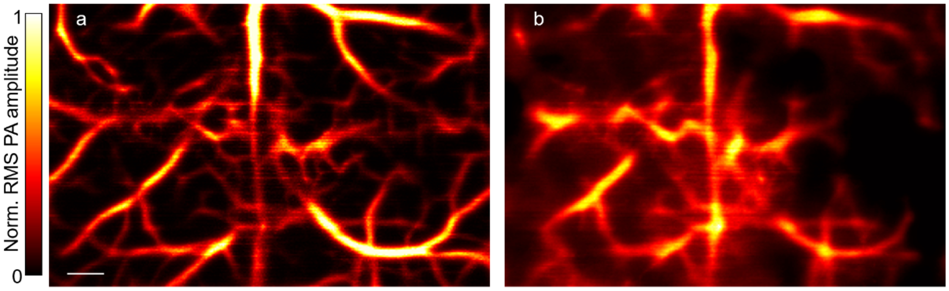
PATER of the mouse cortical vasculature in vivo. (a) Calibration image of mouse brain vasculature. Norm., normalized. Scale bar, 500 μm. (b) Reconstructed widefield image of the calibrated area. All animal procedures were approved by the Institutional Animal Care and Use Committee of California Institute of Technology.