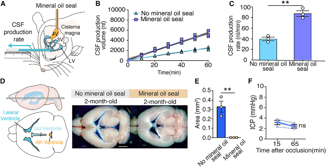
Figure 1. Assessment of the Patency of 4th Ventricular Mineral Oil Seal.
(A) To measure CSF production in mice, a small burr hole was made over the right lateral ventricle (anterior-posterior [AP]: −0.10 mm, medial-lateral [ML]: 0.85 mm from bregma) in C57BL/6J male mice anesthetized with ketamine/xylazine (K/X). A 30-G needle connected to PE-10 tubing was lowered 2.00 mm through the burr hole. The atlantooccipital membrane was surgically exposed and the head rotated 90 degrees downward. A 30-G needle connected to PE-10 tubing filled with mineral oil was inserted into the 4th ventricle, and mineral oil (1 μL, 1 μL/min) was infused, thus occluding the aqueduct of Sylvius. After closure of the aqueduct of Sylvius, CSF was obliged to exit by the needle inserted in the lateral right ventricle. The position of CSF in the PE-10 tube was marked every 10 min for a total of 60 min. The cannula was not inserted in the 4th ventricle in control mice without the mineral oil seal.
(B) CSF production was measured in young male mice under K/X anesthesia with and without injection of mineral oil seal in the 4th ventricle. The best fit lines from the linear regression (colored line) with 95% confidence intervals (shaded region) are plotted. The linear regression in mice without the mineral oil seal is R2 = 0.99, p < 0.0001 and in mice with the mineral oil seal is R2 = 0.99, p < 0.0001.
(C) Comparison of the rates of CSF production with and without the mineral oil seal. CSF production rate is the slope from the linear regression in (B). **p < 0.01, unpaired t test. Bar graphs represent mean ± SEM.
(D) To assess the efficacy of the mineral oil seal on CSF outflow, Evans blue (0.5%, 0.5 μL, 0.5 μL/min, 60-min circulation) was infused into the right lateral ventricle. The brain was harvested, and the distribution of Evans blue was documented by macroscopic imaging. Evans blue remained trapped in the lateral and the 3rd ventricles when the aqueduct of Sylvius was blocked. In contrast, when Evans blue (0.5%, 0.5 μL, 0.5 μL/min, 60-min circulation) was infused in mice with a patent aqueduct of Sylvius, Evans blue was abundantly present in the 4th ventricle (scale bar: 2 mm).
(E) Comparison of Evans blue coverage area in 4th ventricle in mice with (n = 3) or without (n = 3) a mineral oil seal. **p < 0.01, unpaired t test. Bar graphs represent mean ± SEM.
(F) ICP at 15 and 65 min after direct CSF production measurement in mice with mineral oil seal of the 4th ventricle. Paired t test; ns, not significant.

