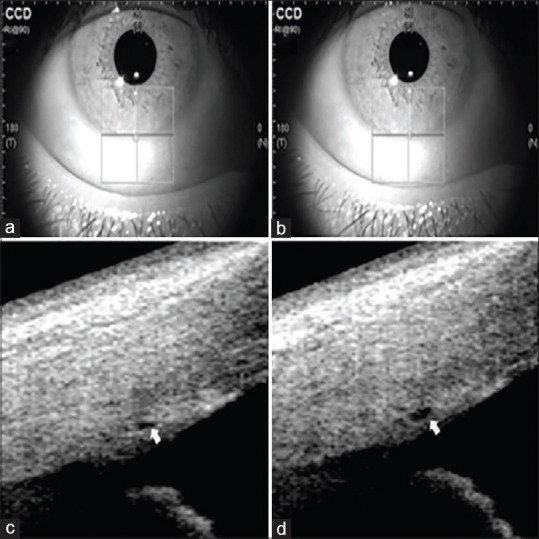
Figure 1.

SS-OCT images of scanning position of SC in POAG eyes. The crypts and furrows of the iris are used to mark the scanning guidelines to ensure the same scanning cross-section of SC before (a) and after (b) aerobic exercise. Morphology of SC before (c) and after (d) aerobic exercise is marked by an arrow
