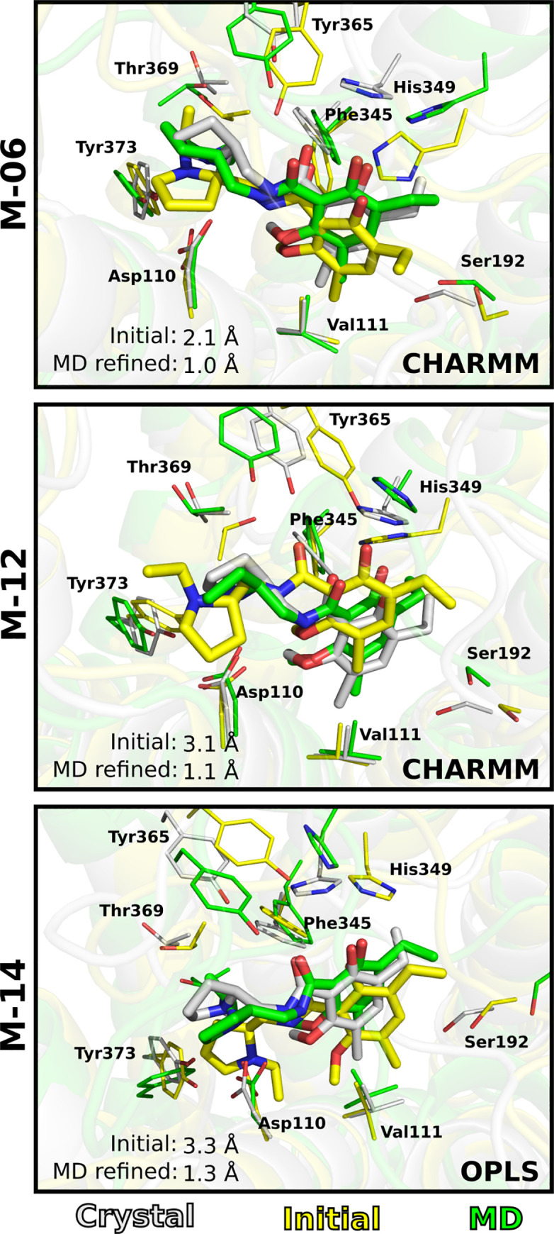
Fig 6. MD refinement of ligand binding mode.

Examples of MD refined binding modes for models M-06, M-12 and M-14. One of the five cluster centroids extracted from the MD simulations is shown. The ligand and key residues are shown as sticks. The initial (yellow), MD refined (green) and crystal (grey) structures are shown as cartoons.
