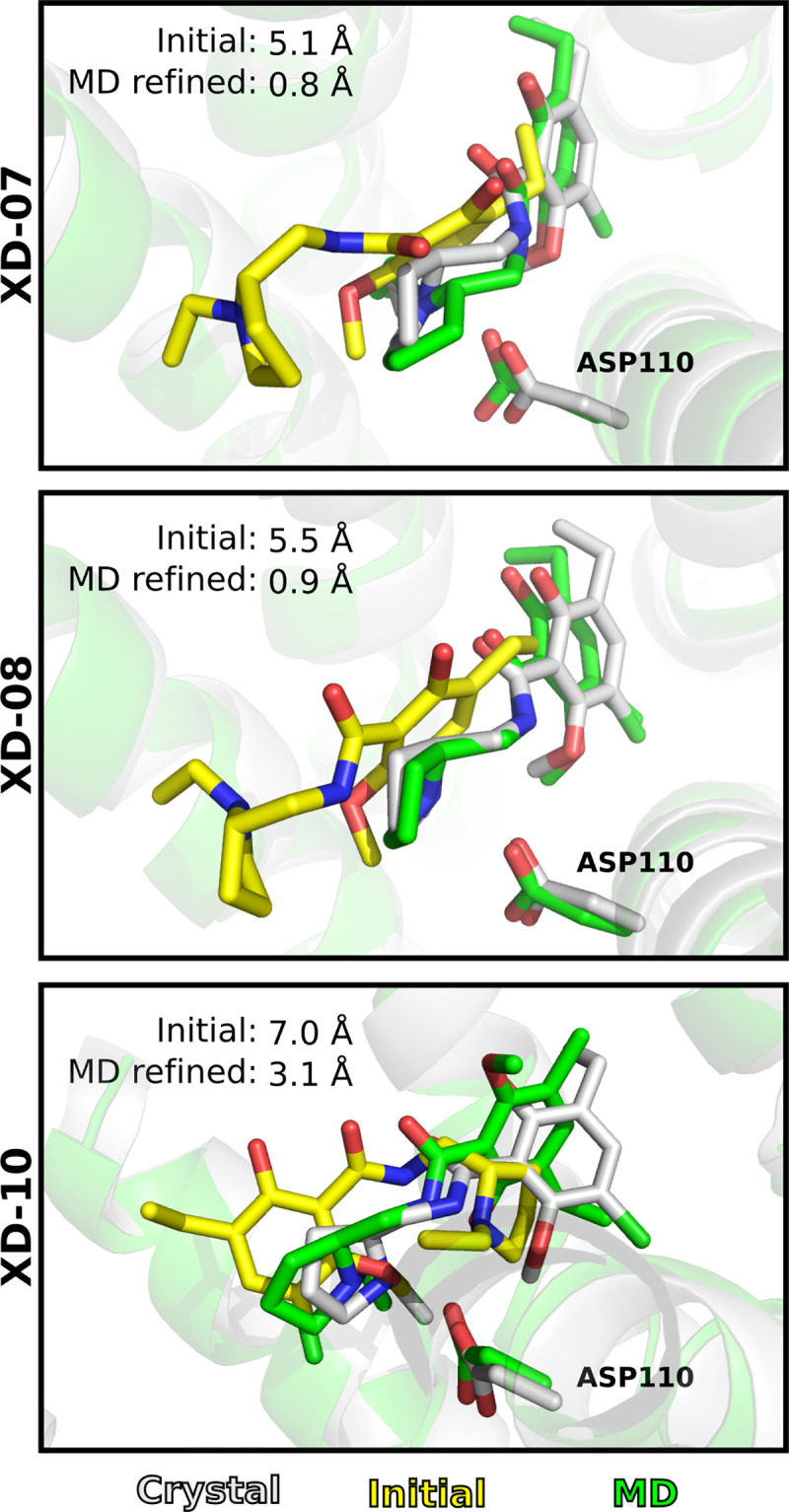
Fig 8. MD refinement of D3R crystal structure with binding modes of eticlopride generated by molecular docking.

The ligand is shown as sticks. The crystal (grey), initial (yellow) and MD refined (green) receptor structures are shown as cartoons. The three models were refined with the CHARMM protocol.
