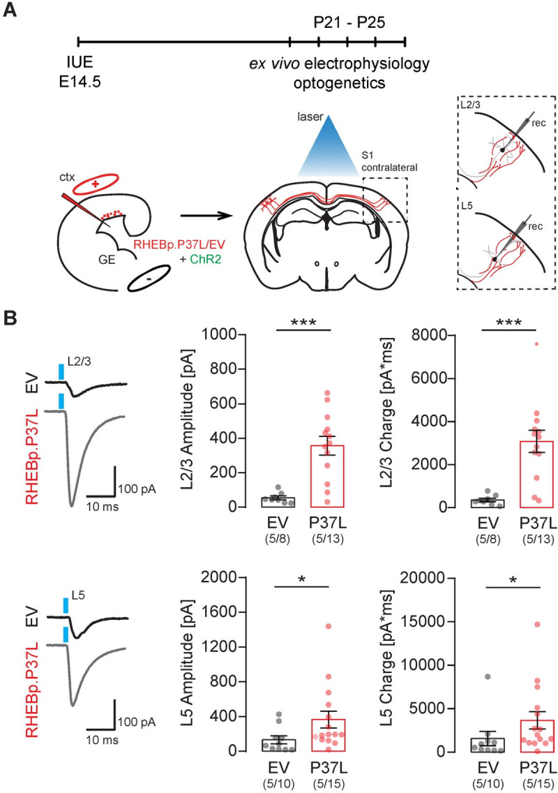
Fig 6. Overexpressing RHEBp.P37L increases synaptic connectivity on the contralateral hemisphere.

(A) Schematic representation of the timeline and experimental conditions of the IUE and ex vivo whole-cell patch clamp recordings in contralateral L2/3 and L5 upon widefield optogenetic stimulation (indicated by the blue laser). (B) Example traces and analysis of the compound postsynaptic responses after photostimulation (blue light), showing the postsynaptic response amplitudes and total charge in contralateral L2/3 and L5 in EV and RHEBp.P37L expressing slices; numbers in the graph indicate number of targeted mice (N = 5) and number of cells (n = 8, n = 13, n = 10, n = 15) analyzed; data are presented as mean ± SEM and single data points indicate the values of each cell; for statistics see S3 Table; the data underlying this figure can be found in S6 Data. *p < 0.05, ***p < 0.001. ctx, cortex; EV, empty vector; GE, ganglion eminence; IUE, in utero electroporation; L2/3, layer 2/3.
