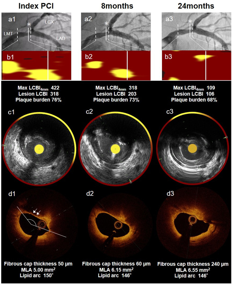
Figure 1.
Case: A 60-year-old-man presenting with unstable angina pectoris. (A1–3) Coronary angiogram reveals mild stenosis in the distal portion of left main trunk artery seen as a non-culprit lesion at index primary percutaneous coronary intervention, 8 months later, and 24 months later. (B1–3) Chemogram at index percutaneous coronary intervention, 8 months later, and 24 months later. (*)minimum lumen area site. (C1–3, D1–3) Near-infrared spectroscopy-intra vascular ultrasound and optical coherence tomography images with minimum lumen area site in non-culprit lesion segment. B1–3 show that yellow pixels were significantly and gradually reduced during the follow-up duration of 24 months. Max LCBI4mm decreased from 422 to 106. The optical coherence tomography at index percutaneous coronary intervention detected a thin-cap fibroatheroma with a thickness of 50 µm (white arrow). This thickness increased to 240 µm at 24 months. UAP, unstable angina pectoris; LMT, left main trunk artery; PCI, percutaneous coronary intervention; OCT, optical coherence tomography; NIRS-IVUS, near-infrared spectroscopy-intra vascular ultrasound; MLA, minimum lumen area.

