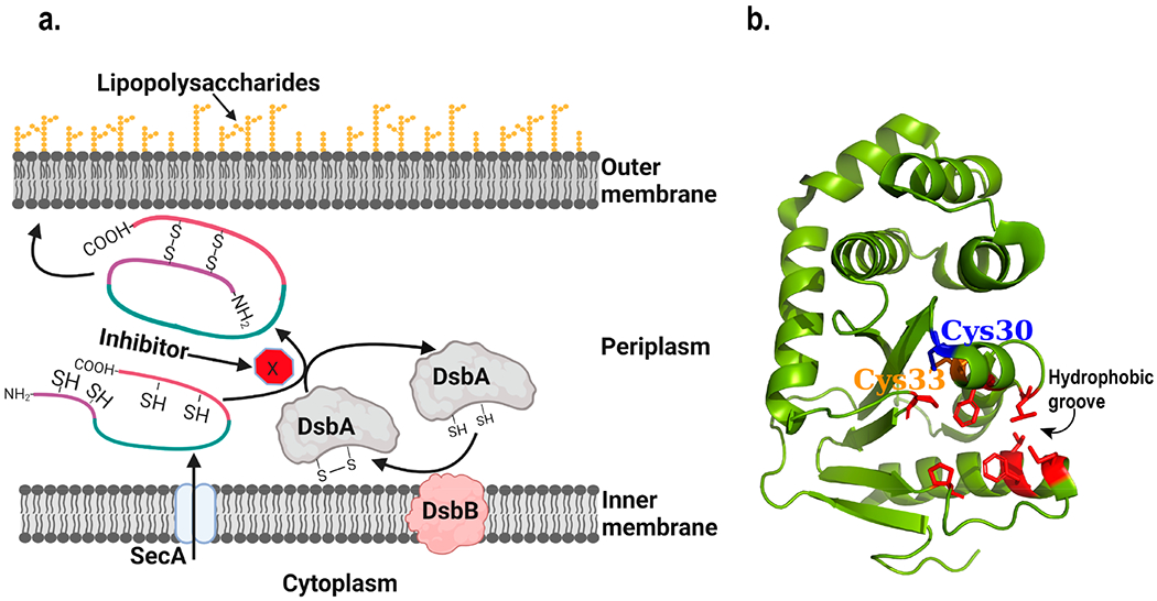
Figure 3.

Structure and function of DsbA. (a) Schematic diagram of the working mechanism of DsbA. The process blocked by inhibitors is highlighted. Figure was created using Biorender. (b) Structure of E. coli DsbA (Created with Pymol73 using 1FVK.pdb). Cysteines present in the active site in the CXXC motif are shown. Residues lining the hydrophobic groove are highlighted in red and with their side chains shown.88
