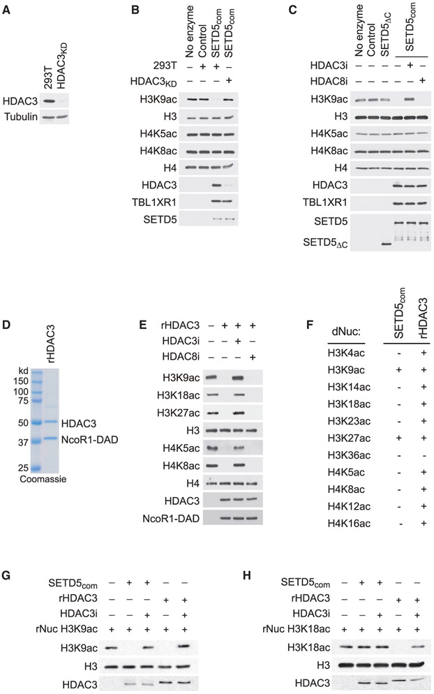
Figure 5. HDAC3-Selective Deacetylation of H3K9Ac by SETD5com.
(A) Generation of HDAC3-depleted 293T cells. Western analysis with the indicated antibodies of control (293T) or HDAC3-depleted 293T cell lysates (HDAC3KD). Tubulin is shown as a loading control.
(B) SETD5com possess HDAC3-dependent lysine deacetylation activity. Western analysis with the indicated antibodies of in vitro histone deacetylation assay on HeLa-purified nucleosomes using SETD5com purified from 293T or HDAC3KD cells.
(C) HDAC3-dependent SETD5com lysine deacetylation activity is inhibited by a selective HDAC3 inhibitor. In vitro deacetylation assays as in (B) ± the selective HDAC3 inhibitor (RGF9966, 1 μM) or ± the selective HDAC8 inhibitor (PCI-34051, 1.5 μM). SETD5ΔC does not interact with HDAC3 (see Figure 3J) and serves as a negative control.
(D) Coomassie stain of active recombinant HDAC3 complex (contains HDAC3 and the DAD domain of NCoR1, labeled as rHDAC3) purified from E. coli.
(E) HDAC3 has broad deacetylation activity on histones. In vitro histone deacetylation assay on HeLa-purified nucleosomes with rHDAC3 complex analyzed by western blots with the indicated antibodies.
(F–H) HDAC3 selectively deacetylates H3K9Ac and H3K27Ac in the context of SETD5com. (F) Summary of deacetylation assays using SETDcom or rHDAC3 on a library of recombinant nucleosomes designed to harbor a single lysine acetylation as indicated. (G and H) Western analysis with the indicated antibodies of deacetylation assays on H3K9Ac rNuc (G) and H3K18Ac rNuc (H). Figure S4 shows the other nine modified nucleosomes summarized in (F).

