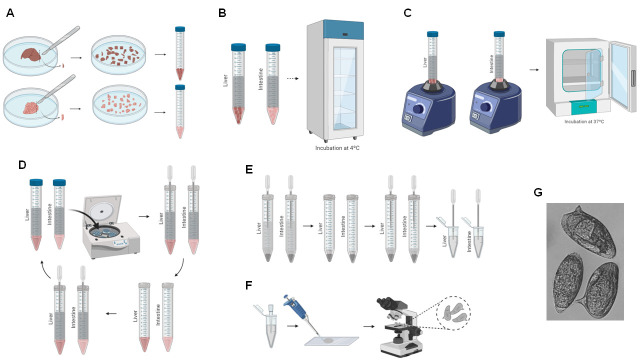
Figure 4. Schema of liver and intestine digestion for egg recovery.
A. Liver and intestine are sliced using a scalpel, and the chopped organs are transferred to a 15-ml Falcon tube (Steps F1-F2). B. 10% KOH is added to the tissue and incubated overnight (Steps F3-F5). C. The tissue is vortexed and incubated at 37°C (Steps F6-F7). D. The 15-ml Falcon tube is centrifuged, the supernatant is removed, and 1 ml 0.85% NaCl is added (Steps F8-F11). E. The resuspended eggs are transferred to a 1.5-ml microfuge tube (Steps F12-F13). F. The recovered eggs are counted (Steps F14-F16). G. Representative image of recovered eggs obtained at 10× magnification.

