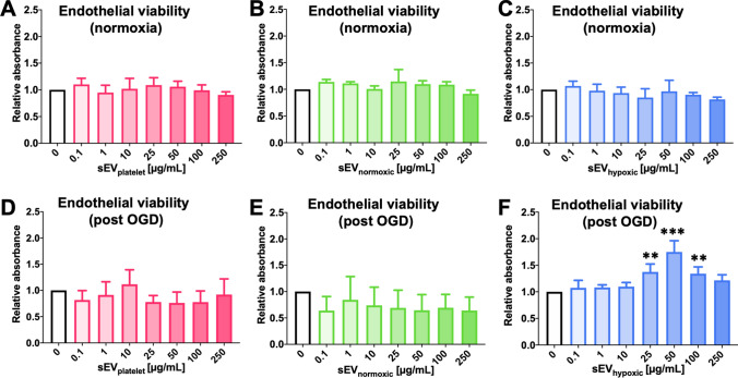
Fig. 4.
sEVs from hypoxic MSCs increase the survival of cerebral microvascular endothelial cells exposed to oxygen–glucose deprivation (OGD), but do not influence the viability of cells cultured under regular ‘normoxic’ conditions. Relative absorbance of hCMEC/D3 cells cultured under (A–C) regular ‘normoxic’ conditions (21% O2) or (D–F) 24 h OGD (1% O2, glucose deprivation) followed by 6 h reoxygenation (21% O2)/glucose recultivation, determined in a 3-(4,5-dimethylthiazol-2-yl)-2,5-diphenyltetrazolium bromide (MTT) assay after exposure to different concentrations of A, D sEVs obtained from MSC culture media that contain platelet lysates (sEVplatelet), B, E sEVs obtained from MSCs (
source 41.5) cultured under regular ‘normoxic’ conditions (21% O2; sEVnormoxic) or C, F sEVs obtained from MSCs (source 41.5) cultured under hypoxic conditions (1% O2; sEVhypoxic). Data are mean ± SD values (n = 3 independent experiments [in A–E], 8 independent experiments [in F]). **p < 0.01, ***p < 0.001 compared with control

