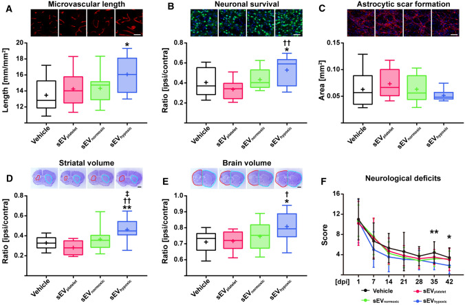
Fig. 6.
sEVs obtained from hypoxic MSCs induce post-ischemic angiogenesis, brain remodeling and neurological recovery in a mouse model of ischemic stroke. A Density of CD31+ cerebral microvessels in the previously ischemic striatum, B number of NeuN+ surviving neurons in the previously ischemic striatum and C area of GFAP+ astrocytic scar in the brain infarct at the rostrocaudal level of the bregma, which is the core of the middle cerebral artery territory, as well as D striatum volume, E whole-brain volume and F neurological deficits evaluated using the Clark score of mice exposed to 40 min middle cerebral artery occlusion (MCAO), which were intravenously treated after 24 h, 72 h and 120 h with vehicle (normal saline), sEVs obtained from MSC culture media that contain platelet lysate (sEVplatelet), sEVs released by MSCs (
source 41.5) cultured under regular ‘normoxic’ conditions (21% O2; sEVnormoxic; equivalent released by 2 × 10⁶ cells) or sEVs released by MSCs (source 41.5) cultured under hypoxic conditions (1% O2; sEVhypoxic; equivalent released by 2 × 10⁶ cells) followed by animal sacrifice after 56 days. Representative microphotographs are also shown. Data are box plots with medians (lines inside boxes)/means (crosses inside boxes) ± IQR (boxes) with minimum/maximum values as whiskers (in A–E) or mean ± SD values (in F) (n = 10 animals vehicle, 6 animals sEVplatelet, 9 animals sEVnormoxic, 9 animals sEVhypoxic). *p < 0.05, **p < 0.01 compared with control/†p < 0.05, ††p < 0.01 compared with sEVplatelet/‡p < 0.05 compared with sEVnormoxic. Scale bars: 50 µm (in A–C)/1 mm (in D, E)

