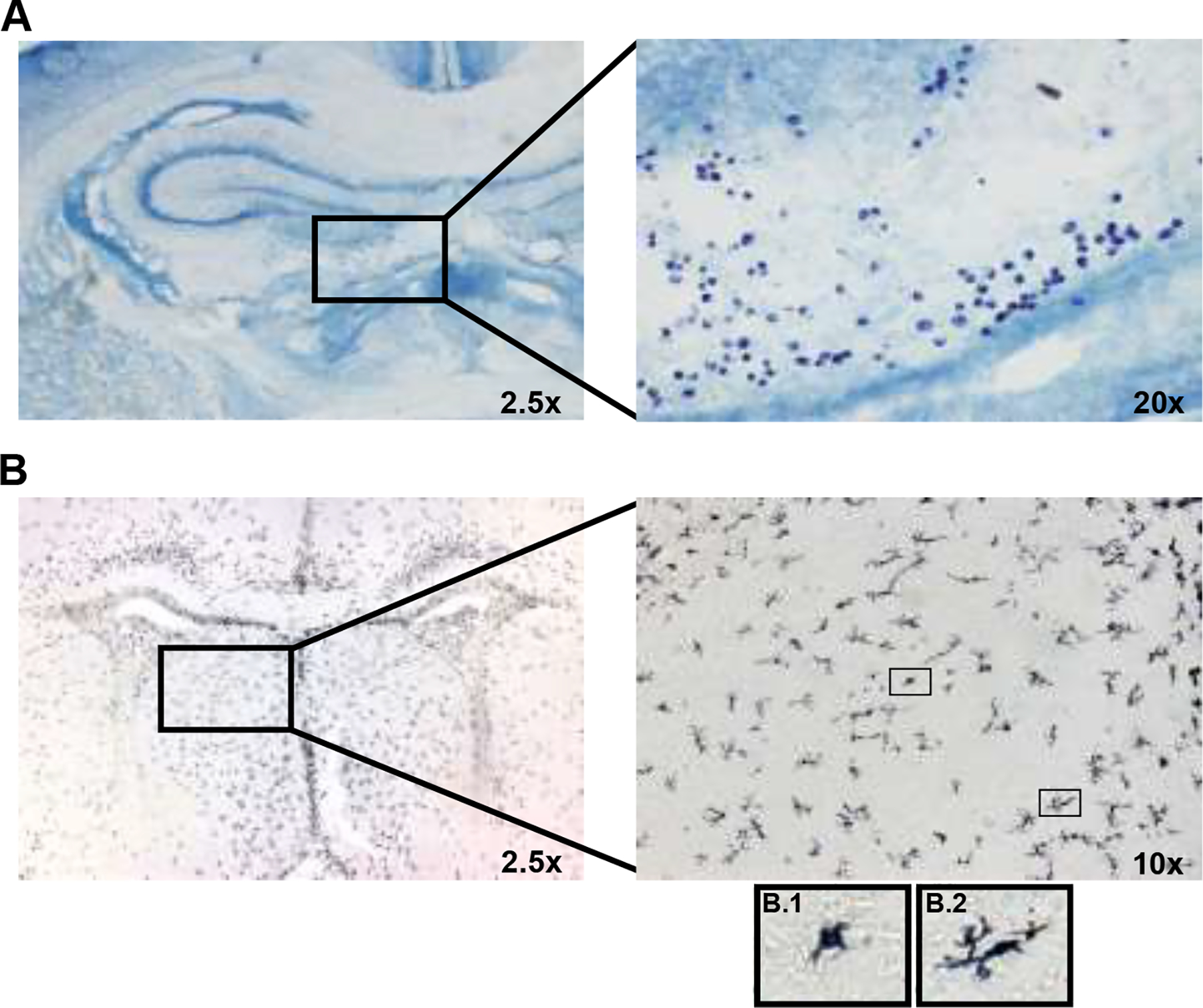
Figure 2.

Representative images of mast cell and microglia staining in a female vehicle offspring on PN0. (A) Toluidine Blue-stained Mast cells in the perivascular area near the hippocampus. (B) DAB-stained Microglia in the lateral septum. Representative ameboid (B.1) and phagocytic (B.2) microglia are pictured.
