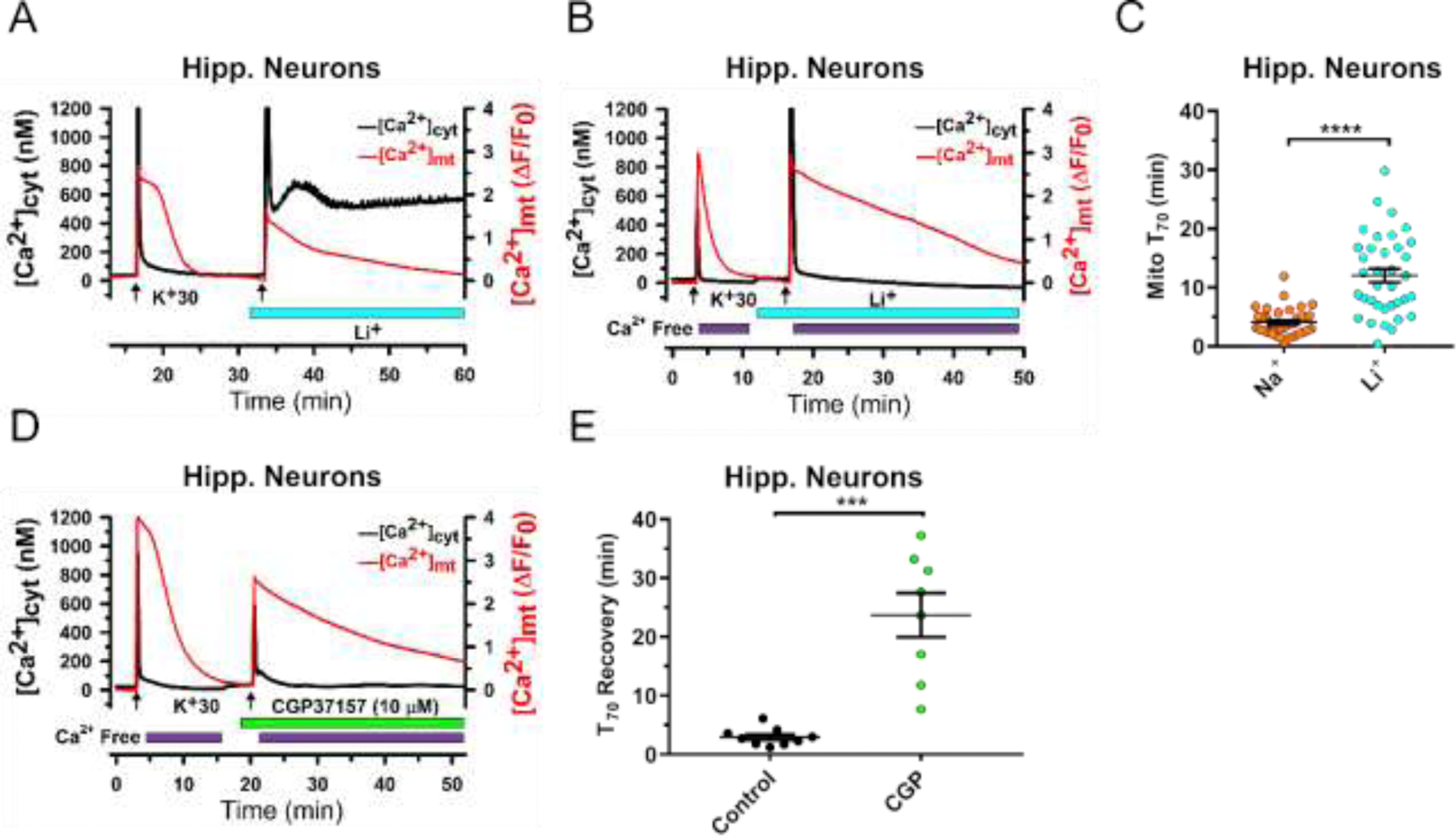
Figure 3. Substitution of Li+ for Na+ markedly inhibits mitochondrial Ca2+ efflux in hippocampal neurons.

(A) and (B), Representative traces from experiments with simultaneous monitoring of [Ca2+]cyt (Fura-2; black) and [Ca2+]mt (mito-RGECO1; red) in cultured hippocampal neurons. [Ca2+]cyt and [Ca2+]mt transients were induced by brief depolarization using 30 mM KCl (K+30, 15 s, arrows). Substitution of Li+ for Na+ in the extracellular solution (0 mM Na+, 140 mM Li+) is indicated by blue bar under the traces. In (B), extracellular Ca2+ was removed (0 mM Ca2+, 100 µM EGTA) immediately after K+30 stimulation (purple bars). (C), Mitochondrial Ca2+ efflux kinetics was quantified by measuring the duration of 70% [Ca2+]mt recovery relative to its peak elevation (T70), and compared between the control conditions (140 mM NaCl in extracellular solution) and following Na+ replacement with Li+ in the extracellular solution. Data are presented as mean ± SEM, n=36; ****p<0.0001, two-tailed paired Student’s t-test. (D) Representative traces of [Ca2+]cyt (black) and [Ca2+]mt (red) show the effect of the mtNCX inhibitor CGP37157 (10 μM; green bar) on mitochondrial Ca2+ efflux in hippocampal neurons. (E), Mitochondrial Ca2+ efflux kinetics was quantified by measuring the duration of 70% [Ca2+]mt recovery relative to its peak elevation (T70), and compared before and after treatment with CGP37157. Data are presented as mean ± SEM, n=9; ***p<0.001, two-tailed paired Student’s t-test.
