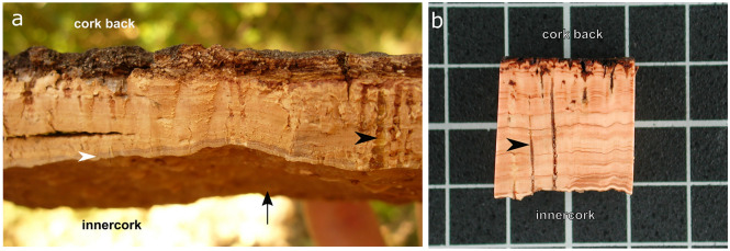
Figure 1.
Anatomy of cork plank (outer bark) from Quercus suber. (a) Cork freshly-harvested at the late spring (June) in Romanyà de la Selva (Girona, Spain) showing this year produced living tissue (white arrowhead) in the innercork side, which faces the phellogen (black arrow). (b) A dried and polished sample is shown to better appreciate the annual cork-rings seen as lighter (earlycork) and darker brown (latecork) zones. Lenticels are highlighted with a black arrowhead. The grid scale distance in B corresponds to 1 cm. The figure was constructed using the free and open source Inkscape software 1.0 (https://inkscape.org/).

