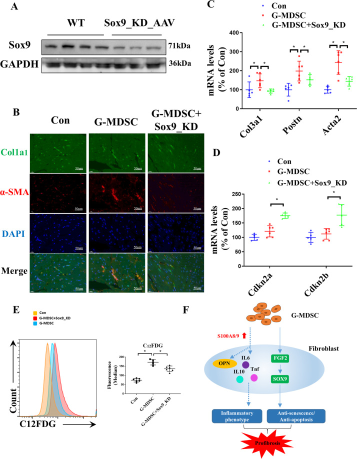
Fig. 7. SOX9 knockdown alleviates the fibrotic phenotypes induced by G-MDSCs in vivo.
Induction of SOX9 knockdown in MDSC-treated mice through injection of AAV harboring the SOX9-KD plasmid. A Western blotting analysis showing the expression levels of SOX9 in the WT and SOX9_KD hearts. B Representative immunofluorescence images of the level of α-SMA in the control, G-MDSC, and SOX9_KD hearts. Scale bars, 50 μm. C The mRNA levels of fibrosis markers (Col3a1, Postn, and Acta2) in mouse hearts analyzed by qPCR; n = 5–6 per group. D The mRNA levels of the senescence markers Cdkn2a and Cdkn2b in mouse hearts analyzed by qPCR; n = 5–6 per group. E The levels of β-galactosidase in fibroblasts analyzed by flow cytometry through C12FDG staining. Representative cytograms are shown on the left, and statistical data are shown on the right; n = 5–6 per group. F Diagram of the mechanism by which G-MDSCs act on fibroblasts. The data are presented as the means ± SDs. Differences were determined by an unpaired t-test or two-way ANOVA (more than 2 factors), and a Sidak HSD post hoc test was performed. *P < 0.05.

