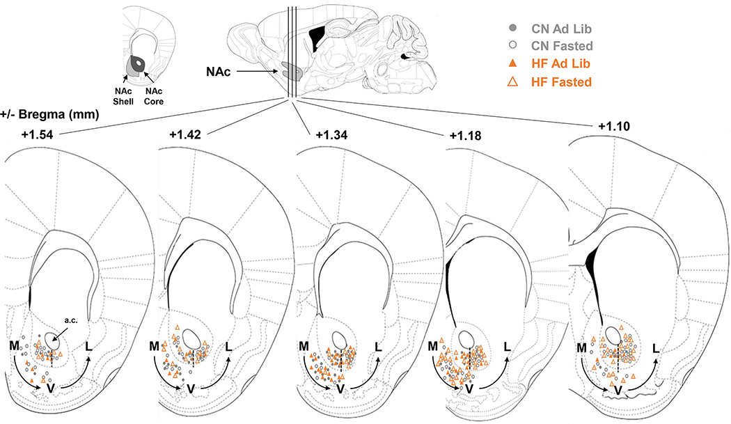
Figure 6: Electrode Placements -.
Recording sites in the NAc core and shell of male and female mice marked at the time of recording and referenced to Franklin & Paxinos mouse brain atlas. Medial (M), ventral (V), and lateral (L) designations are based from the anterior commissure (A.C.), with a dashed line as a landmark for where M→V and V→L groupings were separated.

