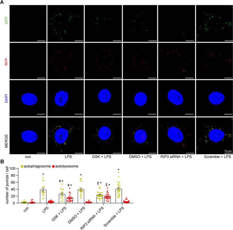
Fig. 4. RIP3 activation suppressed autophagic flux in LPS-induced cultured PTECs.
A, B Cultured PTECs transfected with mRFP-GFP-LC3 adenovirus and treated as indicated for 24 h. Representative confocal laser scanning microscopy images (A) and quantitative analysis (B) of autophagosomes (yellow puncta) and autolysosomes (red-only puncta). Few autophagosomes and autolysosomes were observed in the control group. The number of autophagosomes but not autolysosomes was increased in the LPS-treated PTECs (LPS, LPS + DMSO, and LPS + Scramble group). GSK or RIP3 siRNA induced the formation of autolysosomes, whereas it decreased the accumulation of autophagosomes in LPS-induced PTECs (n = 3–4, 21–26 cells from each group). *P < 0.05, significantly different from the control group. #P < 0.05, significantly different from the DMSO + LPS group. ‡P < 0.05, significantly different from the scramble + LPS group.

