Fig. 4.
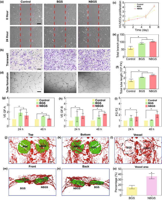
Neovascularization stimulated by BGS and NBGS in vitro and in vivo. a Wound-healing assay using HUVECs cultured with BGS and NBGS for 24 h. b Proliferation of HUVECs cocultured with BGS/NBGS for 1, 3, 5 and 7 days. c Representative photomicrographs of transwell migration assay of HUVECs after 24 h. d Tube formation of HUVECs stimulated by BGS and NBGS for 24 h. e Quantitative analysis of total branch points. f Quantitative analysis of total tube length. g–i Vasculogenesis-related gene expression (VEGF-A, VEGF-B and FGF2) in HUVECs cultured with different scaffolds after 24 and 48 h. j–n Reconstructed 3D micro-CT images of the blood vessels (red) surrounding the scaffolds (green) at 3 weeks. o Quantitative analysis of newborn blood vessels. The scale bar represents 250 μm (a–c) and 1 cm (j–n). (Color figure online)

