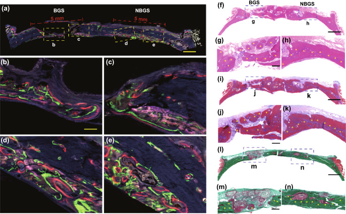
Fig. 6.
Fluorescent imaging and histological staining of the cranial bone defect to assess newborn osseous tissue. a Tetracycline hydrochloride (blue fluorescence), calcein AM (green fluorescence) and alizarin red (red fluorescence) (scale bar, 1 cm) were injected subcutaneously into rats with calvarial defect at week 2, 4 and 6, and different colors of fluorescence represent newborn bone tissue in different durations. Representative newborn bone induced by BGS b, c and NBGS d, e showing the new woven bone around the scaffold at week 8 (scale bar, 250 μm). f–h H&E staining, i–k Masson trichrome staining and l-n Goldner trichrome staining of the cranial defect implanted with BGS/NBGS at week 24. The scale bar equals 2 mm (f, i, l) and 200 μm (g, h, j, k, m, n). (Color figure online)

