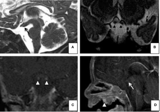
Figure 1.
Magnetic resonance imaging (MRI). Sagittal (A) and coronal (B) T2-weighted images; Gd-enhanced coronal (C) T1- weighted and sagital (D) T1-images show on the midline sagittal plane, a thickened third ventricle floor (arrow a and d), tubomamillary fusion and the absence of a midline sella turcica and pituitary infundibulum. Coronal T2 and T1 WI MR (C) reveal 2 pituitary stalks (arrowheads b and c). The MRI also demonstrates a midline nasopharyngeal teratoma (arrowhead D).

