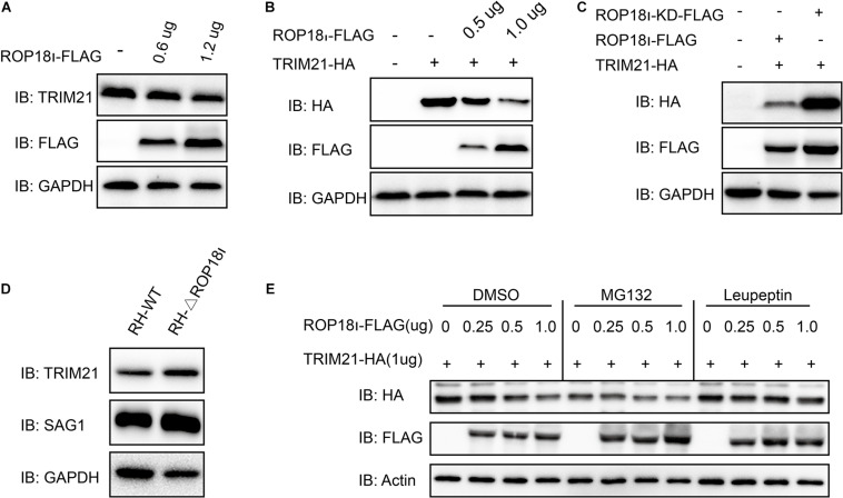
FIGURE 3.
TgROP18I promoted TRIM21 degradation through lysosomal pathway. (A) HEK293T cells were transfected with the increased amounts of pcDNA3.1-ROP18I-FLAG as indicated. (B) HEK293T cells were co-transfected with a stable amount of pcDNA3.1-TRIM21-HA and the increased amounts of pcDNA3.1-ROP18I-FLAG as indicated. The endogenous or overexpressed TRIM21 level was decreased with the increased ROP18 level. (C) HEK293T cells were co-transfected with pcDNA3.1-TRIM21-HA and pcDNA3.1-ROP18I-FLAG or pcDNA3.1-ROP18I-KD-FLAG as indicated. The results of Western blotting detection with the cell lysates indicated that much more TRIM21 was detected in the ROP18-KD overexpression group than in the ROP18 overexpression group. (D) Lysates of HFFs infected with RH or RH-△rop18 was detected by Western blotting, and more TRIM21 was detected in the RH-△rop18 infection group than in the RH infection group. (E) HEK293T cells were co-transfected with 1mg of pcDNA3.1-TRIM21-HA and increased amounts of pcDNA3.1-ROP18I-FLAG. The cells were treated with MG132 or Leupeptin, or left untreated. Cell lysates were subjected to Western blotting, and the results showed that TRIM21’s level was decreased with the increased amount of TgROP18I in the cells treated with MG132 or DMSO. However, TRIM21’s level was kept stable in the Leupeptin treated group. All the experiments were repeated three times. IB, immunoblot.

