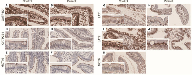
Figure 3.
(A–K) Immunohistochemistry result of ileal LT4 transporters in distal ileum. The panels were taken at × 20 magnification, and inset represents × 40 magnification. OATP1C1 was expressed in both nucleus and brush border in control (A) and the patient (B). OATP2B1 and MCT10 were mainly expressed in brush border in control (C, E) and the patient (D, F), respectively. LAT1 was mainly expressed in brush border in control (G), whereas there was perinuclear staining in the patient (H). LAT2 was detected at both perinuclear area and brush border in control (I), but was stained at only perinuclear area in the patient (J). The expression of MCT8 was not able to check in the patient due to lack of remnant ileal tissue. It was mainly expressed in brush border in control (K).

