
Figure 1.
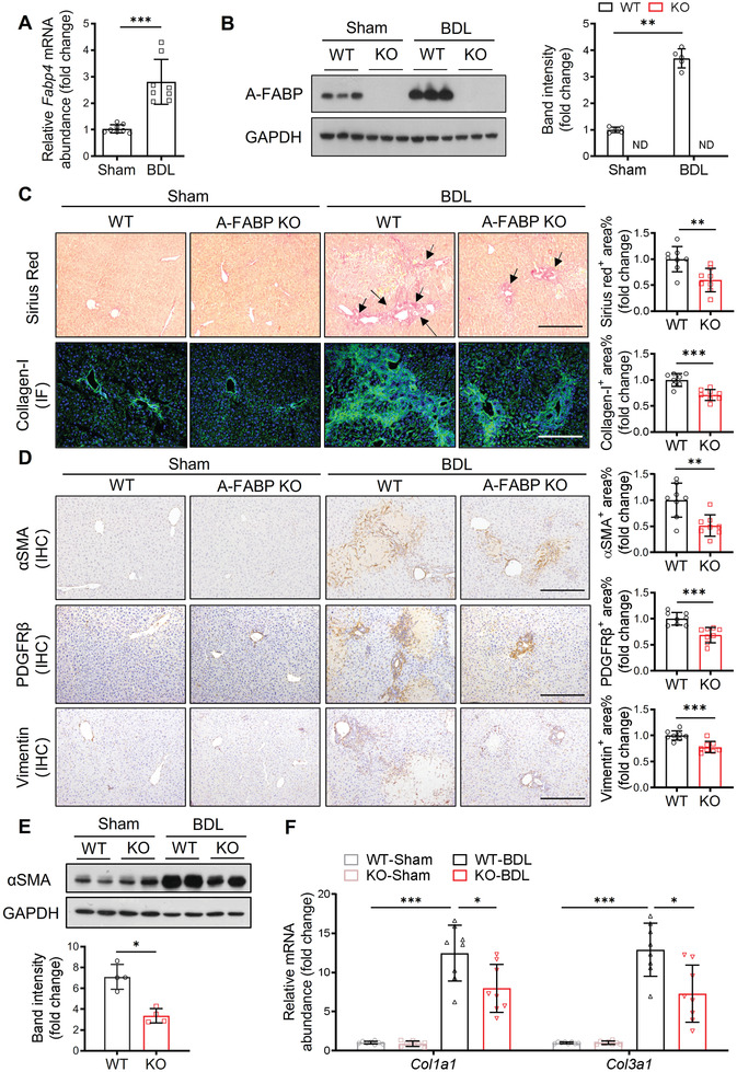
A‐FABP deficiency ameliorates BDL‐induced liver fibrosis in mice. A‐FABP KO mice and their WT littermates were subjected to BDL or sham operation for two weeks. A) Relative mRNA abundance of hepatic Fabp4 in WT mice (n = 8). B) Representative immunoblots of the hepatic expression of A‐FABP and GAPDH in mice and the band intensity of A‐FABP relative to GAPDH (n = 5). Representative images of C) Sirius red staining and immunofluorescence (IF) staining of collagen‐I, and D) immunohistochemistry (IHC) staining of αSMA, PDGFRβ, and vimentin of mouse liver sections (100 ×, scale bar 250 µm) (n = 8). Black arrow in panel C indicates the mature collagen stained by Sirius red. Right panels are the densitometry analysis of the positive area of Sirius red, collagen‐I, αSMA, PDGFRβ, and vimentin of mice in BDL group, respectively (n = 8). E) Representative immunoblots of the hepatic expression of αSMA and GAPDH in mice. Lower panel is the band intensity of αSMA of mice in BDL group relative to GAPDH (n = 4). F) Relative mRNA abundance of hepatic Col1a1 and Col3a1 (n = 8). Data are presented as mean ± SD. *P < 0.05, **P < 0.01, ***P < 0.001. Mann‐Whitney U test was used in (A) and (E). Unpaired Student's t test was used in (B), (C), and (D). Two‐way ANOVA followed by Tukey's test was used in (F).