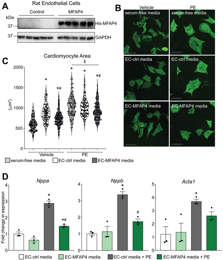
Figure 4. Exogenous endothelial-derived MFAP4 blunts cardiomyocyte hypertrophy.
A. Western blots from rat endothelial cell transfected with GFP control plasmid (Ctrl) or His-tagged MFAP4 (MFAP4) showing MFAP4 expression through His antibodies and GAPDH loading control. B. Representative images of neonatal rat cardiomyocytes treated with either vehicle or phenylephrine (PE), in either serum-free (SF) media, endothelial cell (EC) conditioned media control (EC ctrl media), or conditioned media from EC overexpressing MFAP4 (EC MFAP4 media). Staining shows a-actinin (green). Scale bar, 50 μm. C. Quantification of cardiomyocyte area in the indicated groups (n=100). (Two-way ANOVA results: SF vs EC-Ctrl *p=0.000000000098; SF vs EC-MFAP4 *p=0.0000053; EC-Ctrl vs EC-MFAP4 #p=0.00019; SF vs SF PE *p=0.00000000011; EC-Ctrl vs EC-Ctrl PE *p=0.0000077; EC-MFAP4 vs EC-MFAP4 PE *p=0.0000000055; SF PE vs EC-MFAP4 PE $p=0.0055; EC-Ctrl PE vs EC-MFAP4 PE #p=0.0268). D. qPCR analysis of hypertrophic gene expression relative to Rpl7 in the indicated groups (n=3). (Two-way ANOVA results for - Nppa: EC-Ctrl vs EC-Ctrl PE *p=0.00019; EC-MFAP4 vs EC-MFAP4 PE *p=0.0125; EC-Ctrl PE vs EC-MFAP4 PE #p=0.00096. - Nppb: EC-Ctrl vs EC-Ctrl PE *p=0.00058; EC-Ctrl PE vs EC-MFAP4 PE #p=0.0044. - Acta1: EC-Ctrl vs EC-Ctrl PE *p=0.0435).

