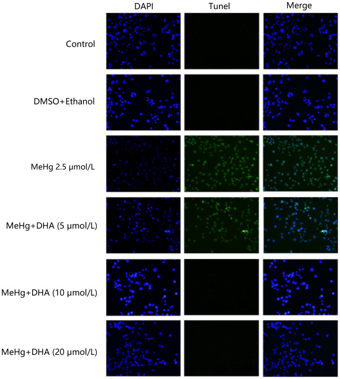
Figure 2.
Effects of DHA on MeHg-induced morphological changes of apoptosis of PC12 cells. TUNEL staining was performed using a TUNEL kit to examine the apoptotic rate of PC12 cells that were pretreated with DHA followed by exposure to MeHg. Blue fluorescence represents the cell nucleus, green fluorescence represents the apoptotic cells (magnification, ×200). MeHg, methylmercury; DHA, docosahexaenoic acid.

