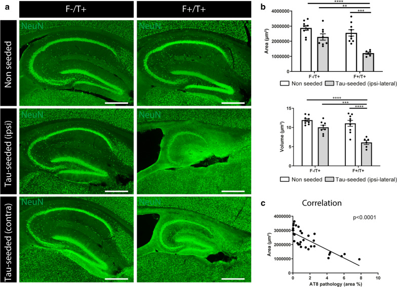
Fig. 2.
Amyloid pathology aggravates tau-induced atrophy. a Representative images of the hippocampus of tau-seeded F−/T+ and F+/T+ mice and their non-seeded littermates at 7 months (3 months post-injection), immunohistochemically stained with anti-NeuN antibody. Scale bar = 500 µm. b Quantification of hippocampal area and hippocampal volume of tau-seeded F+/T+ mice (n = 6) compared to tau-seeded F−/T+ mice (n = 8) and non-seeded F−/T+ and F+/T+ mice (area: n = 9; n = 9). Two-way ANOVA, Tukey’s test for multiple comparison. Data are presented as mean ± SEM; **p < 0.01; ***p < 0.001; ****p < 0.0001 c Correlation analysis between tau pathology in the hippocampus and hippocampal atrophy in 7 months old tau-seeded and non-seeded F−/T+ and F+/T+ mice. Pearson’s correlation analysis

