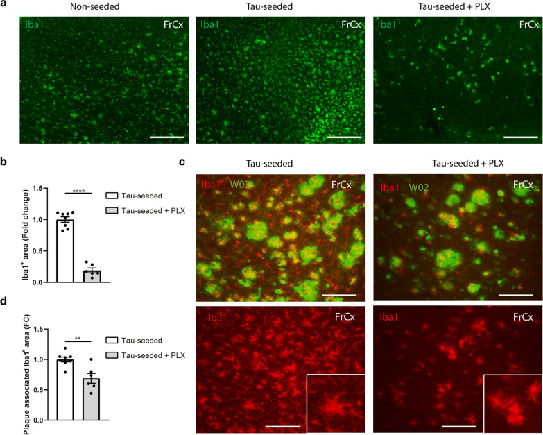
Fig. 6.
CSF1R inhibition preferentially depletes non-plaque associated microglia during ATN pathology. a Representative images of the frontal cortex of tau-seeded F+/T+ mice treated with control, or PLX3397 chow and PBS-injected littermates at 7 months of age (3 months post-injection), immunohistochemically stained with anti-Iba1 antibody. Scale bar = 250 µm. b Quantitative analysis of Iba1 signal in the cortex of PLX3397 treated tau-seeded F+/T+ mice (n = 6) compared to non-treated tau-seeded F+/T+ mice (n = 8). Unpaired t-test. c Representative images of the frontal cortex of tau-seeded F+/T+ mice treated with control or PLX3397 immunohistochemically stained with anti-Aβ antibody W02 and anti-Iba1 antibody and their respective overlay, demonstrating that remaining microglia after PLX3397 treatment reside in the vicinity of amyloid plaques. Scale bar = 100 µm. FrCx = frontal cortex. d Quantitative analysis of Iba1 signal associated with W02+-plaques in the frontal cortex of tau-seeded F+/T+ mice treated PLX3397 (n = 6), non-treated tau-seeded F+/T+ mice (n = 8). Plaque-associated Iba1 signal was quantified as the colocalizing signal and Iba1+ signal immediately bordering W02 positive area. Unpaired t-test. Data are presented as mean ± SEM; **p < 0.01; ****p < 0.0001

