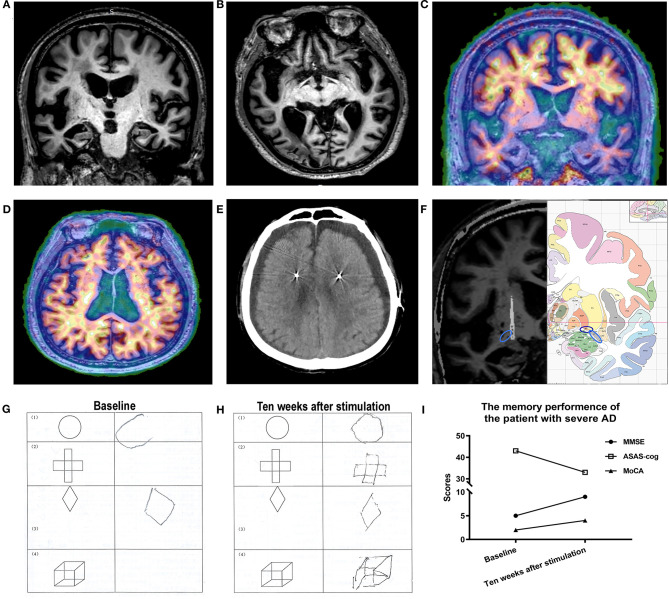
Figure 1.
(A,B) A preoperative MRI revealed global atrophy, especially in the hippocampi and temporal lobes. (C,D) PET imaging showed β amyloid widely deposited in the brain. (E) Bilateral subdural effusion was identified by CT 1 week after surgery. (F) A fused image (left) of the patient's preoperative MRI and postoperative CT showed that the lead (arrow) was accurately implanted into the nucleus basalis of Meynert (NBM) (dark blue circle = NBM; light blue circle = anterior commissure). Atlas (right) of the human brain. (G,H) Patient performance before and after NBM-deep brain stimulation (DBS). The patient was able to draw all geometric shapes after NBM-DBS but performed poorly before NBM-DBS. (I) Changes in memory performance by the patient over time.

