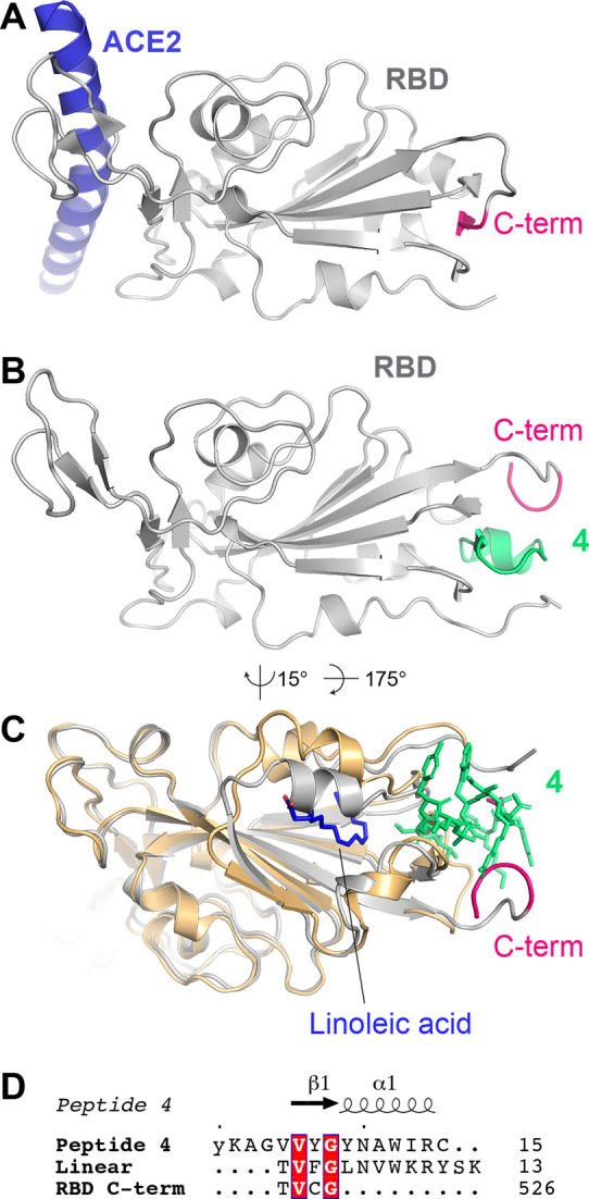
Figure 3.

A) Structure of ACE2 (only helix 1 shown; blue) in complex with the spike RBD (gray; PDB ID 6M0J). The C-terminal β-strand (523TVCG526) is highlighted in pink. B) Structure of the spike RBD in complex with peptide 4 (green). C) Structure of RBD-4 (gray, with 4 shown in green sticks) superimposed onto the spike–linoleic acid complex (PDB code 6ZB4; shown in gold cartoon and blue sticks, respectively). Only the RBD is shown for clarity. D) Alignment of peptide 4, linear RBD-binding peptide ligand,44 and spike RBD C-terminal β-strand. The most C-terminal amino acid shown from the peptide/RBD is numbered on the right.
