
Figure 2.
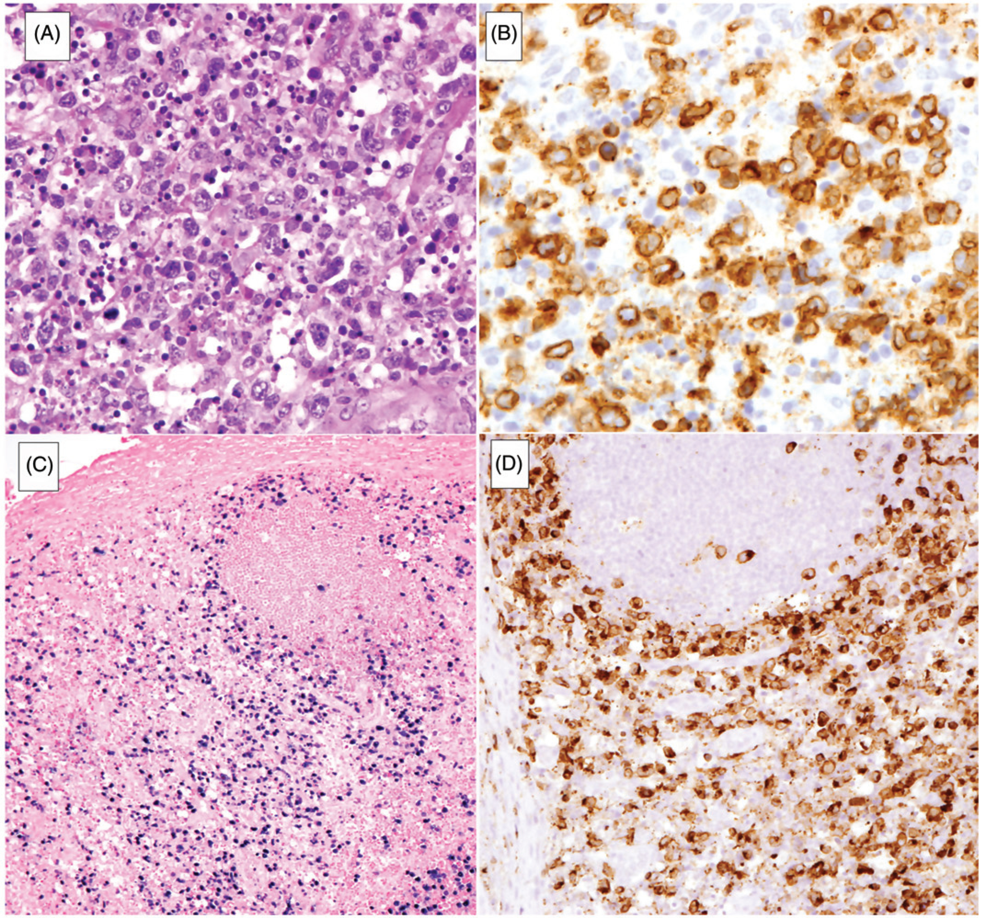
Systemic EBV + T-cell lymphoma of childhood. (A) Lymph node is diffusely infiltrated by atypical lymphoid cells with prominent single-cell necrosis and histiocytic reaction. (B) Atypical lymphoid cells are positive for CD3. (C) EBER in situ hybridization highlights the atypical cells which infiltrate the paracortex, sparing focal reactive follicles. (D) Neoplastic cells are positive for perforin.