
Fig. 4. Increased FasII is responsible for Gef26-associated NMJ morphogenesis.
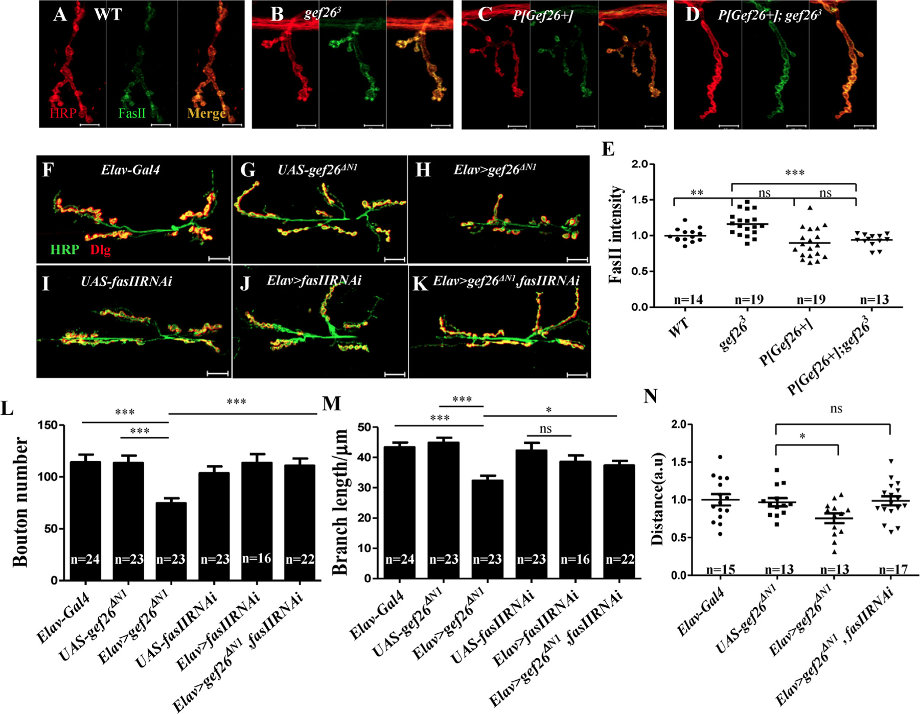
(A-D) Representative images of NMJ4 of third-instar larvae labeled with anti-HRP and anti-FasII antibodies in WT, gef263, P[Gef26+], and the rescue line. Scale bars= 10 µm. (E) Quantification of FasII intensity at NMJ4 in the genotypes indicated in A-D. **p < 0.01; ***p < 0.001; ns, not significant. (F-K) Representative morphology of NMJ 6/7 of the third-instar larvae labeled with anti-HRP and anti-Dlg antibodies in Elav-Gal4, fasIIRNAi driven by Elav-Gal4, and gef26ΔN1 driven by Elav-Gal4 as well as their UAS lines, and Elav-Gal4–driven fasIIRNAi and gef26ΔN1 simultaneously. Scale bars= 20 µm. (L-M) Quantification of bouton number and branch length of NMJ6/7 in the genotypes indicated in F-K. *p < 0.05; ***p < 0.001; ns, not significant. (N) Quantification of a 3-min crawling distance of the third-instar larvae in Elav-Gal4, UAS-gef26ΔN1, Elav-Gal4–driven gef26ΔN1 and Elav-Gal4–driven fasIIRNAi and gef26ΔN1 simultaneously. *p < 0.05; ns, not significant.