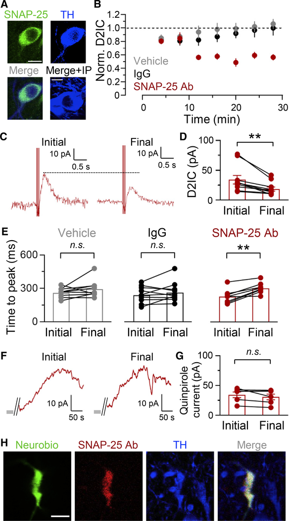
Figure 4. Intracellular application of SNAP-25 Ab decreases evoked D2IC amplitude.
(A) SNAP-25-immunoreactivity (ir) and TH-ir in the SNc, demonstrating SNAP-25 in DA neurons (representative of sections from 18 mice). Pre-adsorption of SNAP-25 Ab with immunogenic peptide (IP) abolished ir (bottom right, sections from 2 mice). Scale bar, 10 μm.
(B) Average time course of changes in evoked D2IC amplitude recorded with single-cell application of SNAP-25 Ab via the pipette (vehicle, n = 16; IgG, n = 12; SNAP-25 Ab, n = 10).
(C) Representative D2ICs immediately after establishing whole-cell recording (initial) and at the end of the recording period (final).
(D) Quantification of D2ICs in the presence of SNAP-25 Ab; mean D2IC amplitude with SNAP-25 Ab was 58% ± 4% of initial, n = 10, **p < 0.01; paired t test.
(E) Time to peak of D2ICs after establishing whole-cell recording and at the end of the recording period. Time to peak (vehicle, initial, 263 ± 13 ms; final, 296 ± 23 ms, n = 9, p = 0.2, IgG, initial, 245 ± 22 ms; final, 267 ± 26 ms, n = 10, p = 0.3, SNAP-25 Ab, initial, 229 ± 21 ms; final, 305 ± 16 ms, n = 8, **p < 0.01).
(F) Representative D2 currents evoked by brief superfusion of quinpirole (250 nM, 15 s). Quinpirole was applied at the beginning and end of recording with SNAP-25 Ab in the pipette. Gray bar indicates quinpirole duration.
(G) Quantification of final quinpirole-evoked D2 currents in the presence of SNAP-25 Ab (90% ± 5% of initial, n = 6, p = 0.2).
(H) Representative immunostaining for SNAP-25 Ab (secondary antibody only) and TH, with Neurobiotin 488 after whole-cell recording; Neurobiotin 488 was introduced with SNAP-25 Ab via the recording pipette (n = 2 slices for triple staining; n = 4 analyzed for Neurobiotin 488 and SNAP-25 Ab). Scale bar, 20 μm.

