Figure 4:

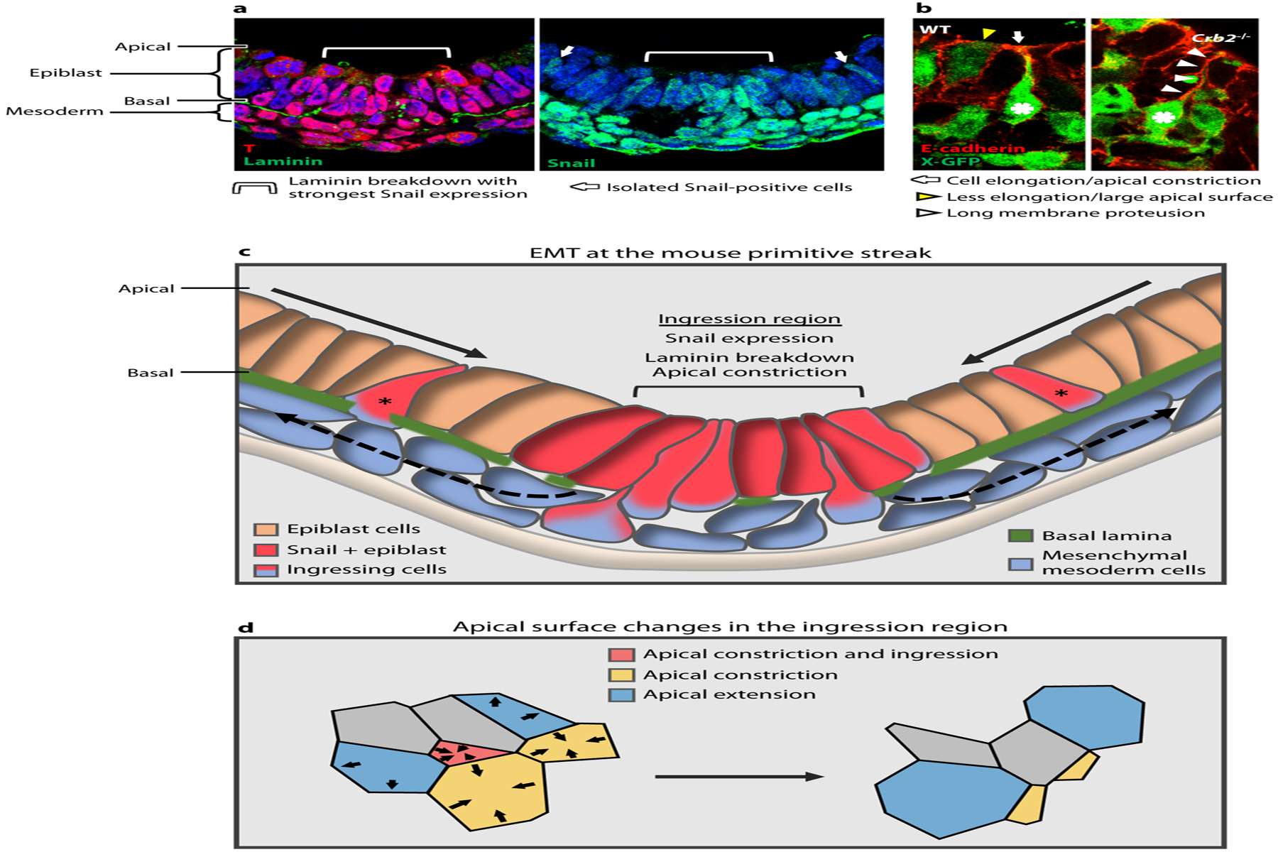
Mouse primitive streak and gastrulation EMT (epithelial-to-mesenchymal transition). (a) Cross-section of the posterior side of a mouse embryo showing T staining at the primitive streak in the epiblast and mesoderm; apical epiblast is up and basal is down. Laminin breakdown occurs beneath a group 6–8 cells wide, corresponding to the strongest Snail expression in the epiblast (light blue brackets). This also corresponds to the region where 80–90% of the ingression events occur, as determined by analyzing whole-embryo time-lapse imaging (data not shown). Isolated Snail positive cells are also observed (white arrows). (b) E-cadherin staining and X-GFP at the primitive streak, show a WT embryo with an ingressing cell (asterisk) elongating and constricting its apical surface (white arrow) and neighboring cell in a different step of the process, which is less elongated and with a larger apical surface (yellow arrowhead). Crb2−/− cell moves further basally (asterisk) and retains a long membrane protrusion attached apically (white arrowheads). Panel adapted with permission from Ramkumar et al. (2016). (c) Schematic of the EMT at the primitive streak during mouse gastrulation. Epiblast cells converge toward the ingression region, which is characterized by expression of Snail, breakdown of the basal lamina, and apical constriction of individual cells. Ingression of a subset of cells at that position appears to be stochastic. Some isolated ingressions are observed outside of this region (asterisks), which may correspond to isolated Snail-positive cells. (d) In the ingression region, cells constrict their apical surfaces in an asynchronous and apparently stochastic manner.
