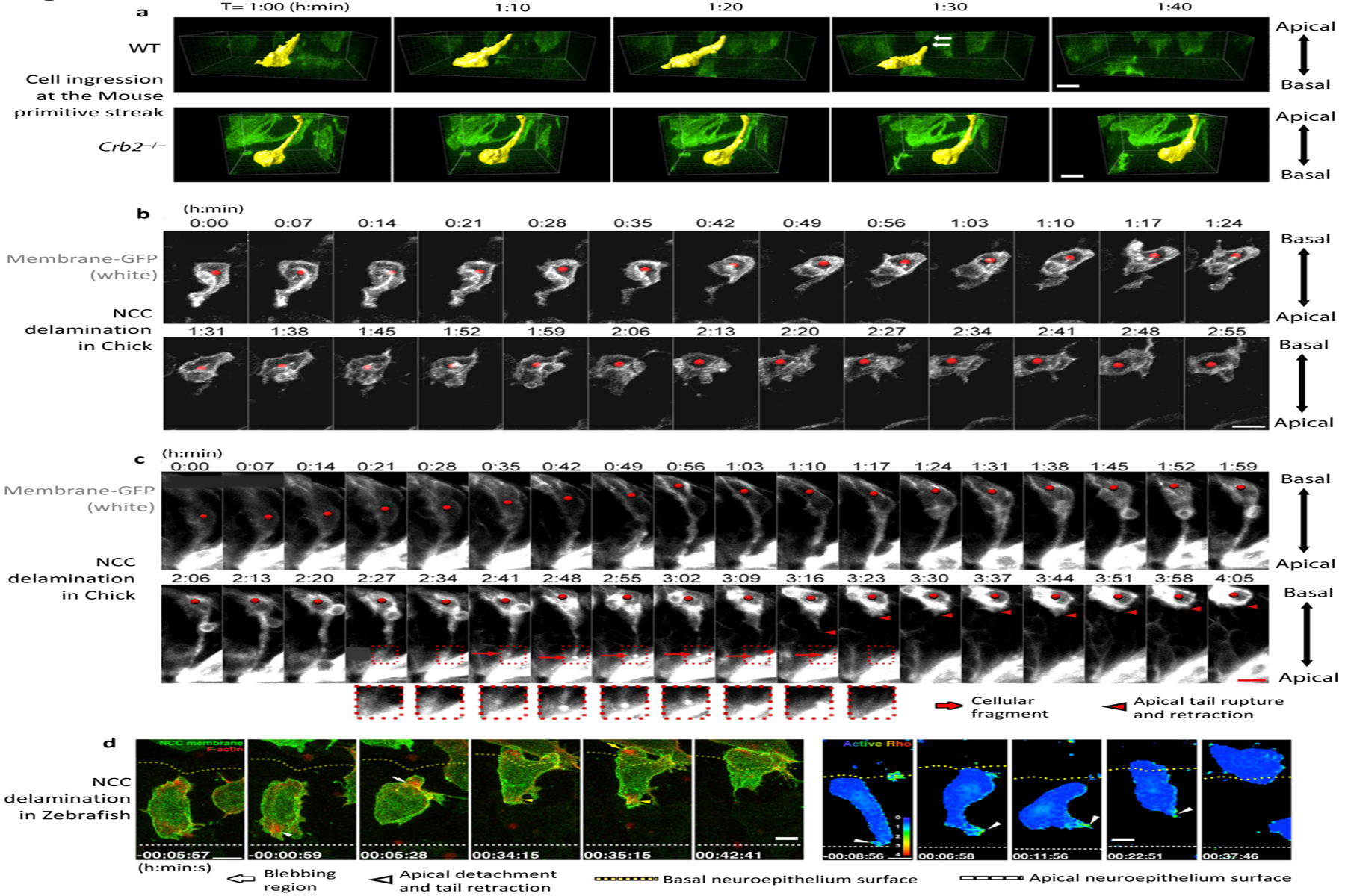
Figure 7:

Dynamic imaging of ingression of cells at the primitive streak and neural crest. (a) Three-dimensional surface rendering of individual cells ingressing at the mouse primitive streak. Wild-type (WT) cells elongate in the basal direction and detach from the apical surface, whereas Crb2−/− cells move basally but remain attached to the apical surface of the epiblast. (b) Dynamics of delamination of Neural Crest Cells (NCC)in the chick embryo, where most cells detach from the apical surface, retract their apical tail, and move out of the epithelium. (c) In some cases, cells detach and leave a fragment of membrane on the apical surface. (d) Dynamics of NCC delamination in zebra fish showing F-actin accumulation in the blebbing region and at the apical tail during apical detachment and retraction, as well as Rho activity in the apical tail during detachment and retraction. In d, apical detachment is at 0 minutes. All scale bars represent 10 μm. Panels adapted with permission from (a) Ramkumar et al. (2016), (b,c) Ahlstrom & Erickson (2009), and (d) Clay & Halloran (2013.
