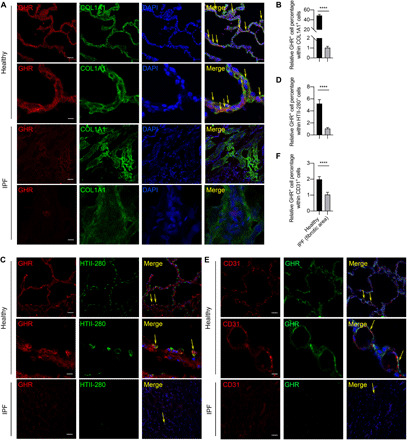
Fig. 2. Most of the mesenchymal cells, and very few ATIIs, are GHR positive in normal lung but not in IPF fibrotic area.

(A) Representative confocal microscopy images of GHR and COL1A1 coimmunostaining for healthy and IPF human lung tissues. (B) Quantification of GHR and COL1A1 coexpression by colocalization calculated from staining in (A). (C) GHR and HTII-280 coimmunostaining reveals that a small portion of HTII-280+ATII cells are GHR positive in normal human lungs and no GHR+HTII-280+ cells in fibrotic human lungs. (D) Quantification of GHR and HTII-280 coexpression by flow cytometry or by colocalization calculated from staining in (C). (E) Rare CD31+GHR+ endothelial cells were found in normal lungs but not IPF lungs. (F) Quantification of GHR and CD31 coexpression by colocalization calculated from staining in (E). Arrows show the coexpressing cells. Scale bars, 100 μm (A to C, lower power images) and 10 μm (A to C, higher power images). Bars represent means ± SEM, n = 7 biological replicates. ****P < 0.0001 by unpaired two-tailed Student’s t test.
