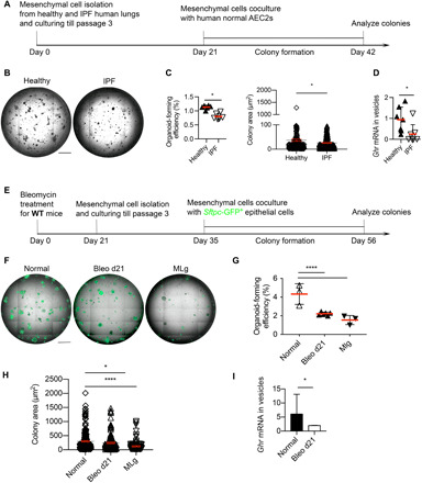
Fig. 7. Fibrotic lung mesenchymal cells retard epithelial regeneration and express less GHR.

(A) Schematic of in vitro coculture human ATII colony formation assay for experiments in (B) and (C). (B) Representative images of ATII organoids cocultured with lung mesenchymal cells from healthy and IPF individuals. (C) Bar graph depicting the organoid-forming efficiency and colony area in (B). (D) GHR mRNA expression in secreted vesicles from cultured lung mesenchymal cells of healthy and IPF individuals (n = 7 in each group). (E) Schematic of in vitro cocultured mouse ATII colony formation assay for experiments in (F) to (H). (F) Representative images of ATII organoids cocultured with mesenchymal cells isolated from normal and bleomycin-treated mice (21 days after treatment), as well as the MLg mouse fibroblast cell line. (G) Organoid-forming efficiency of ATII cocultured with normal, bleo d21, and MLg mesenchymal cells. (H) Colony size of ATII cocultured with normal, bleo d21, and MLg mesenchymal cells. (I) Vesicular expression of Ghr from normal and bleomycin-treated day 21 mouse lung mesenchymal cells cultured in vitro. Scale bars, 1 mm (B and F). *P < 0.05 and ****P < 0.0001 by unpaired two-tailed Student’s t test (C, D, and I) and one-way ANOVA (G and H), means ± SEM.
