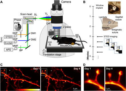
Fig. 1. Repetitive superresolution of the mouse motor cortex using STED microscopy.

(A) Microscope design: A custom-made STED microscope is attached to a microscope stand. The pulsed 483-nm excitation is temporally synchronized electronically with the 595-nm STED light pulses and merged spatially by dichroic mirrors (DM). After passing two galvanic mirrors in the scan-head, the light is imaged by a scan (SL) and tube lens (TL) before being focused by a glycerol immersion objective (numerical aperture, 1.3) with a correction collar. The mouse is mounted via a head bar on an adjustable heating plate. Vital functions are controlled by a pulse oximeter (MouseOx). (B) After window implantation and a 3-week recovery period, the mouse was imaged twice a week. (C) Representative raw data example of an apical dendrite of a pyramidal neuron in the motor cortex of a Thy1-GFP-M mouse imaged at day 1 (left) and day 4 (right). An axon captured in the same field of view is marked by (*). (D) Magnification of marked region in (C). Images are maximum-intensity projections of six frames. APD, avalanche photodiode detector; BP, band-pass filter; MMF, multimode fiber; QW, quarter wave plate; SMF, single-mode fiber. Colorbar, 0 to 212 photon counts. Photo credit: Waja Wegner, University Medical Center Göttingen.
