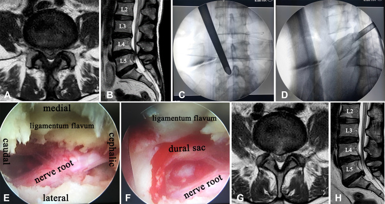
Figure 3.
Pre- and postoperative images of far-migrated LDH. T2-weighted axial MR images of the L4–L5 disk level showing disc extrusion (A) and far down-migrated disc material from the L4–L5 disc level to the L5 lower end plate (B). Intraoperative radiograph showing the placement of the working channel placed between the pedicle and the spinous process (C) and under the pedicle of L5 through limited resection of the lamina and ligamentum flavum (D). Endoscopic view showing complete decompression of L5 nerve root above shoulder and axilla (E). The nucleus pulposus between the dural sac and L5 nerve root was completely removed (F). Postoperative axial magnetic resonance images of the L4–L5 level (G) and T2-weighted sagittal (H).

