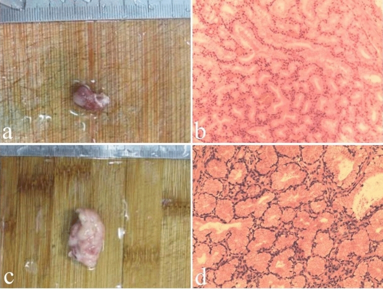
Fig. 3.
Macroscopic and microscopic findings of Brunner’s gland hamartoma. Case 1. A 55-year-old male patient presented with abdominal discomfort and underwent endoscopic examination showing a polyp in the duodenal bulb. a Endoscopically resected specimen of about 1.5 × 0.8 × 0.7 cm in size. b Histological examination revealing massive hyperplasia of Brunner’s glands with focal dysplasia (hematoxylin and eosin, × 100). Case 2. A 60-year-old male patient presented with melena and underwent endoscopic examination showing a polyp in the duodenal bulb with bleeding. c Endoscopically resected specimen of about 3.5 × 2.0 × 1.0 cm in size. d Histological examination revealing massive hyperplasia of Brunner’s glands mixed with smooth muscle and infiltrating inflammatory cells (hematoxylin and eosin, × 100)

