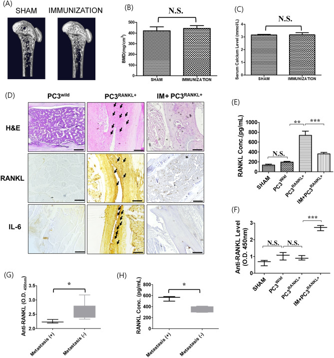
Figure 4.
Effects of immunisation with mutant RANKL on PC3-the innoculated metastatic model. (A) Three-dimensional micro-CT images revealed the trabecular bone architecture of the volume of interest in SHAM and mRANKL-immunized (IMMUNIZATION) mouse femurs (n = 10 images taken in total, one image for each mouse). (B) Bone mineral density (BMD) and (C) Serum calcium Level are shown. No significant differences were observed between two groups. (N.S.) (D) representative H&E stained and immunostained images of RANKL and IL-6 in the PC3Wild, PC3RANKL+ and PC3RANKL+ + IM groups. The metastatic tumor cells or immuno-positive cells were indicated by black arrows. Magnification: × 200. Scale bar = 100 μm. (E) Concentration of RANKL in mouse serum. The mean ± SD values were obtained by densitometry, as shown in the analysis. Significant differences were observed at *p < 0.05 and **p < 0.01 vs. control. (F) Serum samples from mice were obtained after immunisation. Anti-RANKL values in PC3Wild, PC3RANKL+ and PC3RANKL+ + IM groups. The mean ± SD values were obtained by densitometry, as shown in the analysis. Significant differences were observed at *p < 0.05 and **p < 0.01 vs. the control. (G) Anti-RANKL values and (H) RANKL concentration in the serum of RANKL-immunized mice with metastasis (+) or metastasis (−). Bar graphs show the mean ± standard deviation (SD). Significant differences were observed at *p < 0.05, metastasis (+) vs. no metastasis (−).

