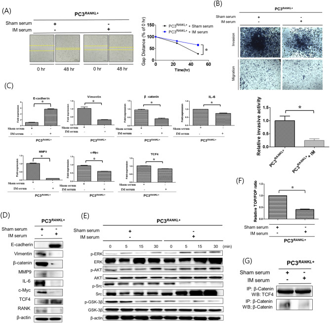
Figure 5.
EMT and metastatic properties of PC3 cells treated with immune sera. (A) Cell migration assays were performed to compare the wound healing capacity of immune serum-treated PC3RANKL+ cells and untreated cells. Immune serum treatment decreased the migration capacity of PC3 cells, as evident from the increase in the wound healing gap at 48 h. Magnification, × 100; scale bar, 100 μm. Data from related graphs are displayed as the mean ± SD. (B) Transwell invasion assays were performed to compare the invasiveness of immunised serum-treated PC3RANKL+ and untreated cells. Serum-treated PC3RANKL+ cells showed a significant decrease in invasiveness. Magnification, × 100; scale bar, 100 μm. Data in the associated graphs are expressed as the mean ± SD. (C) Real-time quantitative polymerase chain reaction for the analysis of EMT and metastasis markers in PC3RANKL+ cells. β-Actin was used as a loading control. Significant differences were observed at *p < 0.05, compared with the control. (D) Expression of E-cadherin, Vimentin, β-catenin, MMP-9, IL-6, c-MYC, TCF-4 and β-actin in immune serum-treated PC3RANKL+ cells, as measured by western blotting. β-Actin was used as a loading control. Similar results were obtained in three independent experiments. (E) Western blot analysis of MAPK phosphorylation levels in serum-treated PC3RANKL+ cells. Similar results were obtained in three independent experiments. (F) TOP/FOP luciferase reporter assays in immune serum-treated PC3RANKL+ cells. Significant differences were observed at *p < 0.05 vs. the control. (G) Co-immunoprecipitation of β-catenin and TCF-4 from PC3RANKL+ and immune serum-treated PC3RANKL+ cells. Each blot was obtained under the same experimental conditions.

