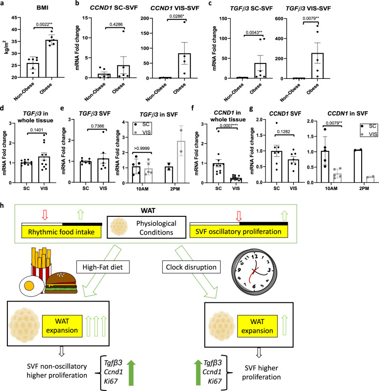
Fig. 8. TGFβ3 and CCND1 expression varies across human adipose tissue depots.
a Body mass index (BMI) from non-obese and obese human patients undergoing prostatectomy intervention (n = 6 patients/group). b, c RT-PCR analysis reveals expression of CCND1 (b) and TGFΒ3 (c) in periprostatic visceral (VIS) or abdominal subcutaneous (SC) SVF prepared from prostatectomy intervention subjects (n = 6 patients/group). mRNA levels from SC fat in lean individuals set to 1. d RT-PCR analysis reveals expression of TGFΒ3 in omental (VIS) and subcutaneous (SC) tissue from obese patients undergoing bariatric surgery (n = 12 patients/fat pad). mRNA levels from the SC fat set to 1. e RT-PCR analysis reveals expression of TGFΒ3 in VIS- and SC-derived SVF (n = 8 patients/fat pad) (left panel) and isolated SVF in the morning (approximately 10:00) (n = 6 patients/fat pad) vs. afternoon (approximately 14:00) (n = 2 patients/fat pad) (right panel) from patients undergoing bariatric surgery. mRNA levels from the SC (left panel) or the SC at 10:00 (right panel) were set to 1. f RT-PCR analysis reveals expression of CCND1 in VIS and SC fat tissue from patients undergoing bariatric surgery (n = 12 patients/fat pad). mRNA levels from the SC fat were set to 1. g RT-PCR analysis reveals expression of CCND1 in VIS and SC isolated SVF (n = 8 patients/fat pad) (left panel) taken at 10:00 (n = 6 patients/fat pad) or 14:00 (n = 2 patients/fat pad) (right panel) from obese patients subjected to bariatric surgery. mRNA levels of all SC (left panel) or the SC at 10:00 (right panel) were set to 1. h Working model of diurnal progenitor proliferation. Data are represented as mean ± SEM. *p < 0.05, **p < 0.01, ***p < 0.001, ****p < 0.0001. Significance (p < 0.05) determined by two-tailed Mann–Whitney U test in (a–c, e, g); two-tailed unpaired t-test in (d, f).

