Figure 2.
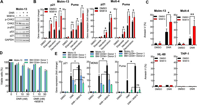
M3814 augments topoisomerase II inhibitor induced p53 apoptotic activity in acute leukemia cells. (A) Molm-13 cells were pre-treated with M3814 (300 nM) or vehicle for 45 min before addition of DNR (10 nM) and incubated for additional 24 h before preparation of protein lysates. Protein expression and phosphorylation status were assessed by Western blotting as in Fig. 1A. Panel displays cropped images from repeat blots divided and probed for different targets. Full-sized blots are shown in Supplementary Fig. 2. (B) Molm-13 and Molt-4 cells were pre-treated with M3814 (300 nM) or vehicle for 45 min before addition of etoposide (50 nM), idarubicin (1 nM) or daunorubicin (10 nM). RNA was isolated 4 h post-treatment and p53 target gene expression was assessed by qPCR using probes for p21 and Puma. Expression levels were normalized to GAPDH. (C) Molm-13, Molt-4, HL-60 and THP-1 cells were pre-treated with M3814 (300 nM) or vehicle for 45 min and exposed to DNR (1 nM). Apoptosis was assessed 24 h post-treatment by flow cytometry as in Fig. 1. D) Molm-13 cells and bone marrow CD34+ cells isolated from two healthy donors were pre-treated with M3814 (300 nM) or vehicle as above for 45 min and DNR (1, 10 or 50 nM) alone or in combination with M3814 (300 nM) was added and the cells were incubated for 4 d. Cell growth/viability was assessed by the CTG assay and expressed as percent of vehicle controls. E) Molm-13 cells and bone marrow CD34+ cells isolated from two independent healthy donors were treated as in D and p53 target gene expression was assessed by qPCR using probes for p21, Mdm2 and Puma. Expression levels were normalized to GAPDH. *p < 0.05, **p < 0.01, ***p < 0.001. NS: p > 0.05.

