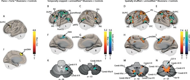
Figure 3.

Neural evidence for decoding communicative intentions in violinists and control participants. (a and j) Increased activity in preSMA and DLPFC for piano vs. forte sequences in violinists vs. control participants. (b,c,f,g) Increased activity for temporally cropped vs. temporally unmodified sequences in violinists vs. control participants in red-to-yellow in IPL, and with overall task performance as group covariates in blue-to-green in IPL, DLPFC, pMTG, preMSA., and cerebellar subregions (k and l). (d,e,h,i) Increased activity for spatially shuffled vs. spatially unmodified sequences for violinists vs. control participants in red-to-yellow in IPS, preSMA, DLPFC, and INS, and with overall task performance as group-level covariates in blue-to-green in IPS, preSMA, DLPFC, INS, and OTC and in cerebellum (m and n). Color bars represent statistical T values of contrast. Black outlines delineate the regions of the group analysis of model 1—not the performance based analysis of model 2. [Cereb: cerebellum lobule; Cereb Crus: cerebellum crus of ansiform lobule; CST: corticospinal tract of the brainstem; DLPFC: dorsolateral prefrontal cortex; IFG: inferior frontal gyrus; INS: insula; IPL: inferior parietal lobule; IPS: inferior parietal sulcus; lingual gyrus; OTC: occipito-temporal cortex; pMTG: medial temporal gyrus; posterior part; preSMA: pre supplementary motor area; PostCG: postcentral gyrus; RO: Rolandic operculum; SupraMargG: supra marginal gyrus; Ver: vermis; Violon.: violinists; Con.: control participants; L: left; R:right]. Voxel-wise P < 0.05 FDR corrected.
