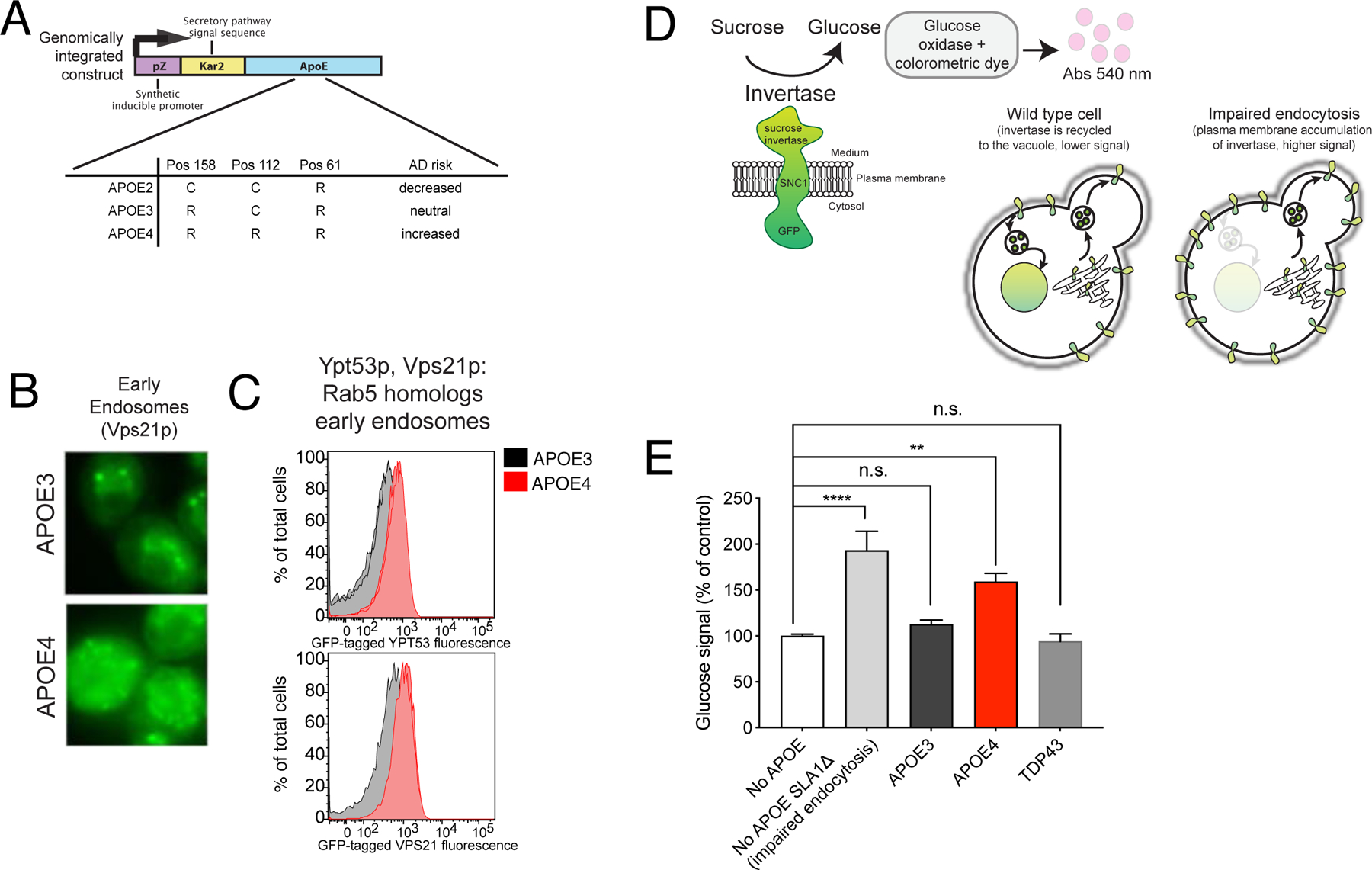
Figure 2. A yeast model faithfully recapitulates APOE4-associated endocytic defects.

(A) The estradiol-inducible promoter system used in the APOE yeast model. (B) Fluorescence microscopy of the mNeonGreen-tagged Vps21p. (C) Mean fluorescent signal from mNeonGreen-tagged Vps21p and Ypt53p quantified by flow cytometry, 10,000 cells per sample. (D) A schematic of the GSS invertase assay adapted. (E) Glucose signal from invertase assays of the indicated APOE-expressing strains and a TDP43-expressing strain. One-way ANOVA followed by Tukey’s multiple comparisons test (**** p-value<0.0001). Mean ± SD for n=3 independent colonies. See also Figure S3.
