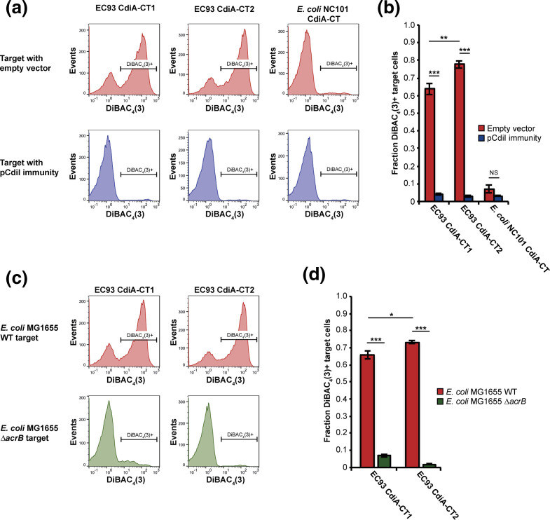
Fig. 4.
The CdiA-CT1 and CdiA-CT2 toxins are membrane ionophores that require AcrB for activity. Membrane depolarization analyses using DiBAC4(3) (see Materials and Methods). E. coli MG1655 inhibitor cells expressing the CdiA-CT1, CdiA-CT2 or CdiA-CTNC101 toxins (see Materials and Methods), depicted at the top of the panels, were mixed with E. coli MG1655 target cells in M9Glu+casAA liquid medium, then analysed by flow cytometry. In the lower three panels, targets expressing cognate CdiI immunity proteins were analysed. (b) Quantification of the data in (a) as the fraction of target cells that were depolarized (n=6 biological replicates ±sem). (c) AcrB-dependence of membrane depolarization. Analysis was carried out as in (a) using E. coli delivering the EC93 CdiA-CT1 or CdiA-CT2 toxins (top of panels) and wild-type or ΔacrB target cells (left of panels). (d) Quantification of the data in (c) as the fraction of target cells that were depolarized (n=4 biological replicates ±sem). Statistical significance was determined by two-tailed, unpaired t-test (*P<0.05, **P<0.005, ***P<0.0005).

