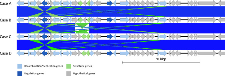
Fig. 4.
Easyfig alignment of prophage 2 in all cases order descending A–D, detailing a small inversion from samples A and B relative to samples C and D (light blue). Arrow indicates gene direction. Recombination/replication genes shown in light blue, regulation associated genes in dark blue. Structure and lysis associated genes shown in light and dark green respectively, finally grey are hypothetical genes.

