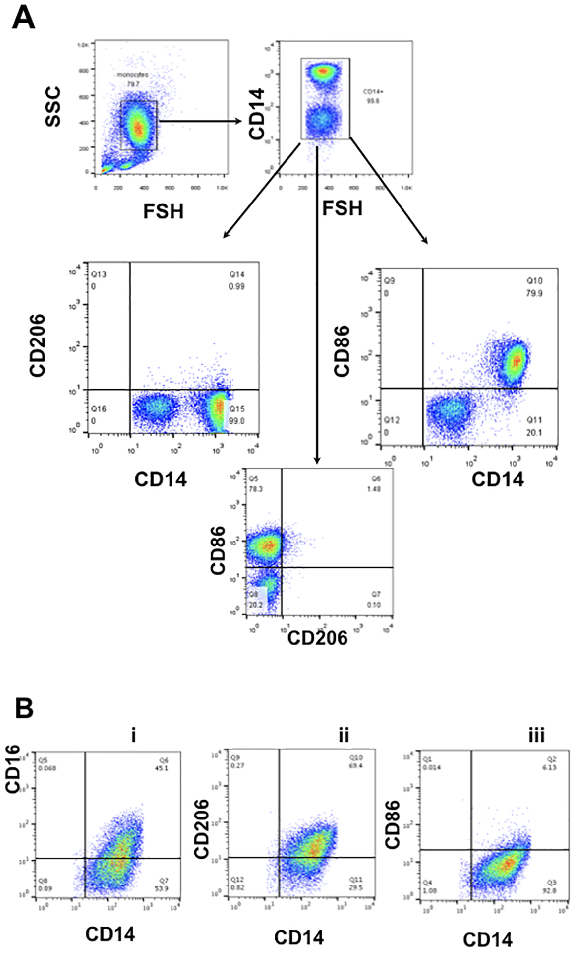
Figure 1. Phenotype of peripheral blood monocytes before and after differentiation stimulated by Trophoblast CM.

A. Gating strategy was set up for characterization of circulating monocytes. Isolated peripheral blood CD14+ monocytes express CD14 and CD86 but not CD206 (CD14+/CD206−/CD86+)
B. Freshly isolated CD14+ monocytes were exposed to first trimester trophoblast CM for 6 days and then analyzed by flow cytometry for CD14, CD86, CD206 and CD16. Following treatment with trophoblast CM, monocytes/macrophages maintained CD14 expression, and gained CD16 and CD206, but lost CD86 (CD14+/CD16+/CD206+ /CD86−). Representative figure of 6 independent experiments with each done in triplicate.
