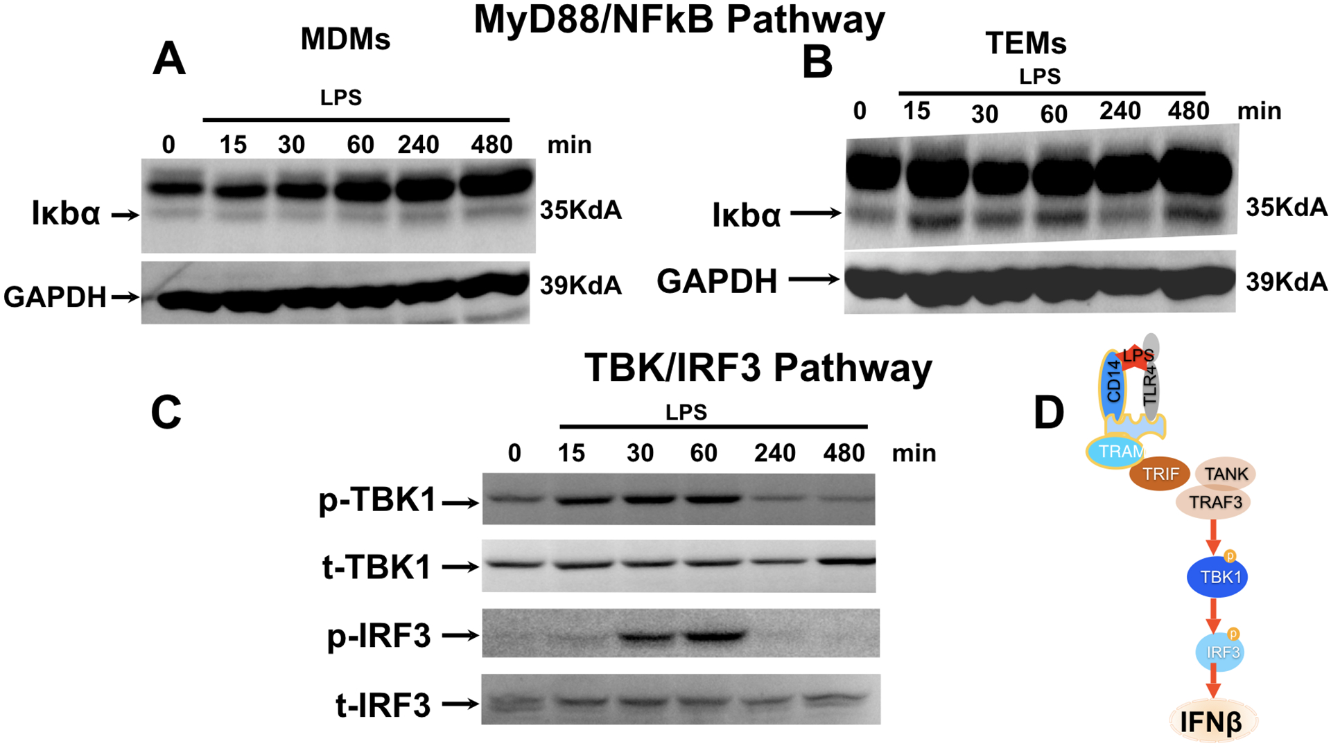
Figure 5. LPS-TLR4 ligation in TEMs is associated with the activation of the MyD88 independent pathway TBK/IRF3.

Freshly isolated CD14+ monocytes were differentiated with trophoblast CM (TEM) or M-CSF (MDM) for 6 days followed by treatment with LPS (10 ng/ml) for 15, 30, 60, 240, and 480 mins.
A. Effect of LPS treatment on MyD88/NFκB pathway was determined in M-CSF polarized macrophages (MDMs) by analyzing levels of IκBα by Western blot analysis. Note the low levels of IκBα expression in MDM.
B. Effect of LPS treatment on MyD88/NFκB pathway was determined in trophoblast educated macrophages (TEMs) by analyzing levels of IκBα by Western blot analysis. Note the continue high expression of IκBα in the presence of LPS treatment.
C. Effect of LPS treatment on TBK/IRF3 pathway was determined by analyzing phosphorylation status of TBK and IRF3 by western blot analysis. pTBK1=phosphorylated TBK; tTBK1=total TBK1. pIRF3= phosphorylated IRF3; t-IRF3=total IRF3. Representative figure of 3 independent experiments.
